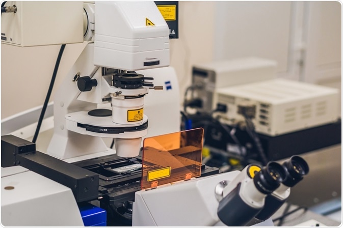
Confocal microscopy, often dubbed as laser confocal scanning microscopy (LCSM), is an imaging technique that allows for 3D capturing with a good resolution. This is accomplished through the construction of a micrograph, derived from high-intensity laser light phased through the pinhole of a digital camera.
This apparatus was created to circumvent the problems of fluorescence microscopy, which uses high-intensity UV light. This intense light brought about by the mercury arc lamps inside fluorescence microscopes could cause disturbing background noise and can even lead to photobleaching.

Image Credit: Elizaveta Galitckaia/Shutterstock.com
The confocal apparatus
Images in a confocal microscope are taken with a digital camera fitted with a pinhole, a staple feature amongst these devices. This guides the light from only one focal plane to be focused through the digital camera, while light from above or below its fulcrum is simply negated.
The laser that is fixed to the device focuses on select regions of a sample in succession, meanwhile, the fluorescent dye that is adsorbed by the sample is captured by the digital camera. Two rotating mirrors found within the apparatus allow for a series of images to be taken.
By accruing this sequence of images from different focal planes (analogous to stacking image layers), one can compile a full 3d image using software of choice. By exploiting this method of 3d imaging, marginal improvement can be denoted on both the axial (z-axis) and lateral (x and y-axis) planes. These renditions of cells and other matrices are made possible by illuminating the object using a double inverted cone of light.
One issue in using standard epi-fluorescence microscopes is that thicker matrices/specimens will exhibit a greater degree of fluorescence. The emission given off from samples that are greater than 2 micrometers will jeopardize the veracity of the generated image, losing fine detail. Confocal microscopes address this by narrowing the excitation ranges while using optical sectioning to eliminate background noise.
Applied confocal microscopy
One case study performed by Donghee Lee et al. highlights one practical application of confocal microscopy. The extracellular matrix (ECM), or the non-cellular components that pertain to all cells, is imperative in cell function. The elasticity of the ECM regulates many biological functions of the cell, such as differentiation of mesenchymal stem cells, adhesion, and migration.
To properly diagnose, treat, and elucidate these tissues in vitro, hydrogels of tunable elasticity are used for mechanobiology studies. These tests are performed because Young’s modulus of hydrogel substrate mimics the ECM appropriately.
Confocal microscopy has been used in conjunction with this indentation method applied to polyacrylamide gels. This automated image process method can successfully measure these indentations and perforations in the ECM mimic. Therefore, confocal laser microscopy successfully gauges the depth and rigidity of PAAM gel compositions, effectively expounding upon a direct mimic to the ever-critical ECM. This has provided an attractive alternative to the AFM nanoindentation method and microneedle method.
This is easily accomplished because confocal imaging can reliably probe anywhere between 0-200 µm beneath the surface of most specimens. It is important to note, however, that each specimen has its own specified depth at which scattering, photobleaching, and noise are too high. It is said that there are some specimens where confocal microscopy should be avoided altogether, as they would no longer be valid.
Introduction to Confocal Microscopy
Superlative/alternative forms of microscopy
Two-photon microscopy, or multi-photon microscopy, generates two photons to accomplish the work of one. When acquiring two photons, each with half energy, exiting in quick succession, this process will depend on the square of the intensity of light hitting the sample.
The infrared lasers that pulse at an order of around 100 MHz will make out of focus light much dimmer than in-focus light. This means that the only excitation that will occur is at the focal spot of the microscope. This will effectively limit the excitation volume, which means that no pinhole will be required, juxtaposing confocal microscopy.
This technique, though similar in nature, has amassed some popularity- as more and more are beginning to discover its advantages. The scattered light from two-photon microscopy does not excite fluorescence, because the infrared waves it uses will scatter less than high-intensity laser light. This will minimize signal loss and allow for the penetration of tissues that are roughly 1 mm, surpassing confocal microscopy’s capacity to penetrate 200 μm tissue. The lower energy infrared light will also minimize the damage done to cells.
One advantage that confocal microscopy does hold against two-photon microscopy, is its superior resolution. A general rule of thumb regarding electron microscopes is that the resolution is inversely proportional to the wavelength (λ). This implies that confocal microscopes (with their higher energy and smaller wavelength) will have a superior resolution when rivaling two-photon microscopes (with their lower energy and longer wavelength.)
Many advancements have been in the field of microscopy since the unveiling of the electron microscope in 1931. Though confocal microscopy is considerably more powerful than conventional widefield techniques or fluorescence microscopy, two-photon microscopy and transmission electron microscopes have become more commonplace. Confocal microscopy then embodies the bridge between these methodologies.
Sources:
- Donghee Lee, Md. Mahmudur Rahman, You Zhou, and Sangjin Ryu. (2015) Three-Dimensional Confocal Microscopy Indentation Method for Hydrogel Elasticity Measurement, Langmuir 31 (35), 9684-9693
- W. Göhde, U. C. Fischer, H. Fuchs, J. Tittel, Th. Basché, Ch. Bräuchle, A. Herrmann, and K. Müllen. (1998) Fluorescence Blinking and Photobleaching of Single Terrylenediimide Molecules Studied with a Confocal Microscope, The Journal of Physical Chemistry, 102 (46), 9109-9116
- (1996) Analytical Currents: Measuring fluorescence decay with a confocal microscope, Analytical Chemistry, 68 (1), 16A-16A
- Webb, D. J., & Brown, C. M. (2013). Epi-fluorescence microscopy. Methods in molecular biology (Clifton, N.J.), 931, 29–59.
- Benninger, R. K., & Piston, D. W. (2013). Two-photon excitation microscopy for the study of living cells and tissues. Current protocols in cell biology, Chapter 4, Unit–4.11.24. https://doi.org/10.1002/0471143030.cb0411s59
- Zhongming Li, Kyle Aleshire, Masaru Kuno, and Gregory V. Hartland. (2017) Super-Resolution Far-Field Infrared Imaging by Photothermal Heterodyne Imaging, 121 (37), 8838-8846
Further Reading