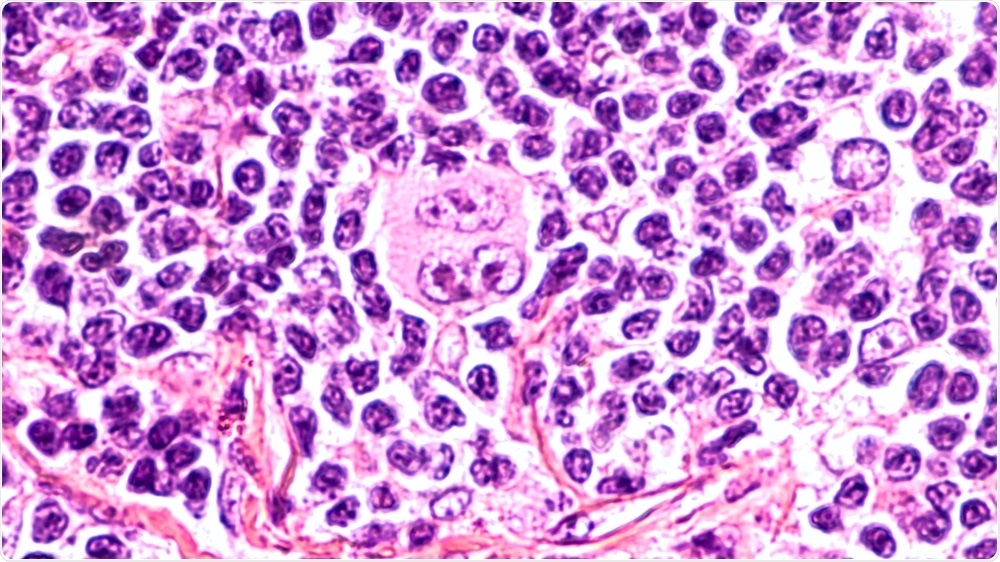
Scientists have discovered significant differences in the neighborhood preferences of Hodgkin lymphoma tumor cells compared with inflamed lymph nodes that could help develop computational methods for analyzing and diagnosing cases of Hodgkin lymphoma.

Image Credit: David. A. Litman/Shutterstock.com
Current diagnostic methods hinge on human error
A team across two institutions in Germany, the Johann Wolfgang Goethe-University and Technische Hochschule Mittelhessen, designed a study to investigate the distribution of tumor cells and their microenvironment in Hodgkin lymphoma, to find significant patterns of behavior that could be used to help flag cancerous cells.
The German team recognized that there is a need for new computational methods that can quantify and standardize tissue images collected from potentially pathological sites.
Currently, in pathology, the analysis of tissue images is conducted by trained pathologists who view and evaluate the samples with a light microscope.
While pathologists are extensively trained to recognize the signs of pathology in tissue, the method is still limited by human skill and at risk of human error.
To enhance the accuracy and reliability of tissue image analysis, scientists aimed to develop a computational method that would be able to quantify and standardize histological observations.
Using the microenvironment to determine diagnosis
The diagnosis and monitoring of disease progression and treatment effectiveness can benefit from histopathology, where tissues are examined under a microscope by histopathologists who make educated interpretations based on their training and experience.
However, the process of histological diagnosis remains challenging, notably, for tumorous diseases, it can be difficult. Because of this, there is a demand for a standardized technique to be used alongside histopathologist assessments.
Therefore, the German team embarked on a research project to quantify experimental data to elucidate markers of disease that could be used to support or histopathological assessments.
A tumor’s microenvironment has become of particular interest in recent years, with a growing body of research demonstrating that the area surrounding tumorous cells is significantly different from that of healthy cells, meaning that it can be used for diagnosis.
The present study specifically looked at the analysis of Hodgkin lymphoma tissue, systematically exploring tumor cell properties as well as their spatial neighborhood relations. To do this, the team studied over 400,000 immunohistochemically stained whole slide images of tissue samples taken from cases of classical Hodgkin lymphoma, nodular sclerosis, and mixed cellularity, and lymphadenitis (inflammation of the lymph nodes).
A statistical analysis was conducted, comparing the differences in properties and microenvironment between these distinct case types.
Results showed that certain morphologies of cells were linked with specific favored and unfavored spatial neighborhood relations, meaning that these neighborhood preferences could potentially be used as indicators of disease.
The researchers believe that these key differences could help distinguish between Hodgkin lymph nodes containing tumor cells, and inflamed lymph nodes, two scenarios that can look similar under the microscope. The findings showed that CD30-positive cells in the microenvironment are different in different diagnoses.
Future prospects
A new approach was established for the analysis of tissue images to help determine the presence of tumorous cells, and well as to determine the spread of these cells throughout the tissue.
The team highlighted significant neighborhood preferences that can be used to distinguish between pathological cells and healthy ones.
The team believes that their new approach could be easily modified to apply to different types of tumor, offering a simple and reliable method to be used alongside conventional histopathological methods to improve tumor diagnosis.
In the near future we could see this method being developed for use in clinical settings, although it is unlikely to replace traditional techniques, rather, it would be used to complement them.
Journal reference:
Hannig, J., Schäfer, H., Ackermann, J., Hebel, M., Schäfer, T., Döring, C., Hartmann, S., Hansmann, M. and Koch, I. (2020). Bioinformatics analysis of whole slide images reveals significant neighborhood preferences of tumor cells in Hodgkin lymphoma. PLOS Computational Biology, 16(1), p.e1007516.