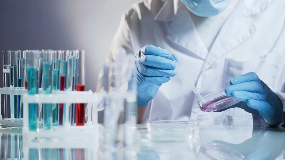
3D organoid models have revolutionized toxicology, providing a more accurate representation of in vivo conditions. These three-dimensional structures mimic human organ tissue, allowing researchers to assess the toxic effects of various substances with precision. This innovation offers great promise for safer, more efficient drug development and toxicity testing.

Image Credit: Motortion Films/Shutterstock.com
What are 3D Organoid Models?
3D organoid models are miniature, simplified versions of organs created in vitro that replicate the key functional, structural, and biological complexities of actual organs.1 These models are derived from stem cells, including embryonic stem cells, induced pluripotent stem cells (iPSC), and adult stem cells (adSC), which can self-organize in a three-dimensional culture due to their self-renewal and differentiation capacities.1
Organoids are complex systems, as they can develop intricate interactions between multiple cell types, closely emulating the structure and function of real organs like the lung or heart.1
They are typically grown in a supportive matrix, which enables cells to thrive and interact naturally in three dimensions.1 This environment is crucial for the cells to form organ-like functions and for the organoids to be used for sustained growth.1
These models have a wide range of applications in biomedical research, including drug testing, personalized and regenerative medicine, gene repair, transplantation therapy and cancer research.1
Advantages Over Traditional Methods Used in Toxicology Testing
3D organoids offer a more physiologically relevant model for toxicology testing compared to traditional 2D cultures, as they can replicate the complex structure and function of human tissues.2
These organ-like structures enable high-throughput screening and more sensitive detection of toxic compounds, potentially identifying hazards before widespread human exposure.2 Organoids also provide a platform for evaluating tissue-specific toxicities, which is crucial for accurate predictions of human responses.2
Unlike traditional models, organoids maintain the genetic and phenotypic heterogeneity of the source tissue, which is essential for studying the effects of toxic substances.2 They can be used for drug screening with high efficiency and sensitivity, guiding personalized treatment strategies.2
Organoids also reduce the need for animal testing, addressing ethical and financial concerns.2 They can be derived from patient-specific cells, enhancing the relevance of the model for personalized medicine.2
Applications in Toxicology Research
3D organoids serve as valuable tools for modeling the adverse effects of drugs, potentially leading to safer pharmaceuticals. They have been successfully generated for nephrotoxicity screening, particularly, human kidney organoids have been used to confirm the nephrotoxic effects of drugs like cisplatin.3 This is crucial in identifying potential kidney damage before the drugs are administered to patients.
Organoids are also being employed to screen for genetic susceptibility to conditions like non-alcoholic fatty liver disease (NAFLD) and non-alcoholic steatohepatitis (NASH), providing insights into genetic risk factors.4 They have proven effective in testing the impact of 247 enzyme inhibitors against diseases like autosomal dominant polycystic kidney disease (ADPKD)5.
Furthermore, human hepatic organoids have been developed to replicate metabolism and CYP450-mediated drug toxicity, offering a solution for drug metabolism assays.6 This is indispensable in predicting how drugs will be metabolized in the human body and identifying potential toxicities early in the drug development process.6
In precision oncology and personalized medicine, organoids derived from patient cells are being used to develop patient-specific models, enabling the most effective treatment for an individual to be determined by testing potential drugs on experimental models derived from patient samples.7
Lastly, human liver organoids offer a sensitive method for liver toxicity screening.8 They have detected that certain drug combinations, safe individually, can be toxic to the liver when combined, potentially preventing severe liver damage in patients.8
Challenges and Limitations
While a significant advancement in biomedical research, organoid technology encounters a critical limitation due to the absence of a vascular system.9 As organoids increase in size, the inner cells suffer from a lack of nutrients and waste accumulation, affecting their growth and functionality.9
Efforts to induce vascularization include in vivo methods, where organoids are implanted into animal models to develop blood vessels, and in vitro techniques, such as gene editing and co-culturing with endothelial cells.9
However, these approaches face challenges: in vivo methods may lead to the replacement of organoid structures with animal components, while in vitro strategies often result in incomplete and non-functional vascular networks.9 These limitations underscore the need for the development of new models, such as organ-on-a-chip.9
3D organoids are also limited by their inability to fully replicate the immune environment found in living organisms.9 Traditional 2D cultures and organoids can be co-cultured with immune cells, but these setups fall short of emulating the complex interactions between immune cells and other tissue cells.9
This limitation is particularly evident in the field of tumor immunology, where the immune microenvironment plays a critical role in cancer progression and the response to drugs.9 Although some progress has been made in creating organoids that include immune components, these models do not yet accurately represent the intricate tumor-immune interactions.9
The current methods still struggle to maintain the unique tumor-immune microenvironment over time.9 This represents a significant barrier to the use of organoids in studying immune responses.9

Image Credit: Motortion Films/Shutterstock.com
Regulatory Implications in 3D Organoids for Toxicology Testing
Despite the promise of organoids, there are regulatory challenges that must be addressed to fully integrate them into the drug development pipeline.10 The production of organoids for clinical applications must adhere to strict quality standards, including good manufacturing practices (GMP), to ensure reproducibility, proper cellular composition, and functional activity.10
The regulatory framework for organoids is still evolving, and their use in clinical settings will need to meet the stringent requirements for cell and gene-based therapies.10 Advances in bioengineering, such as 3D bioprinting and vascularization, may enhance the scalability and robustness of organoid production, which is crucial for their translation into effective medical applications.10
Future Directions
The future of toxicology testing is poised to be revolutionized by using 3D organoids, which offer a more physiologically and human-specific model than traditional 2D cultures and animal models.2
Organoids have the potential to bridge the translational gap between preclinical studies and human trials by more accurately mimicking human tissues.2 This is particularly important for drug development, where organoids can be used to predict drug efficacy and toxicity, thereby improving the efficiency of the drug discovery process and potentially reducing the reliance on animal studies.2
Sources:
- Zhao Z, et al. (2022). Organoids. Nature Reviews Methods Primers, 2(1). https://doi.org/10.1038/s43586-022-00174-y
- Kim J, et al. (2020). Human organoids: model systems for human biology and medicine. Nature Reviews Molecular Cell Biology, 21(10), 571–584. https://doi.org/10.1038/s41580-020-0259-3
- Digby J. L, et al. (2020). Evaluation of cisplatin-induced injury in human kidney organoids. American Journal of Physiology-Renal Physiology, 318(4), F971–F978. https://doi.org/10.1152/ajprenal.00597.2019
- Han D. W, et al.(2023). Customized liver organoids as an advanced in vitro modeling and drug discovery platform for non-alcoholic fatty liver diseases. International Journal of Biological Sciences, 19(11), 3595–3613. https://doi.org/10.7150/ijbs.85145
- Tran T, et al. (2022). A scalable organoid model of human autosomal dominant polycystic kidney disease for disease mechanism and drug discovery. Cell Stem Cell, 29(7), 1083-1101.e7. https://doi.org/10.1016/j.stem.2022.06.005
- Kim H, et al. (2022). Development of human pluripotent stem cell-derived hepatic organoids as an alternative model for drug safety assessment. Biomaterials, 286, 121575. https://doi.org/10.1016/j.biomaterials.2022.121575
- Dao V, et al. (2022). Immune organoids: from tumor modeling to precision oncology. Trends in Cancer, 8(10), 870–880. https://doi.org/10.1016/j.trecan.2022.06.001
- Zhang C. J, et al. (2023). A human liver organoid screening platform for DILI risk prediction. Journal of Hepatology, 78(5), 998–1006. https://doi.org/10.1016/j.jhep.2023.01.019
- Huang Y, et al. (2021). Research Progress, Challenges, and Breakthroughs of Organoids as Disease Models. Frontiers in Cell and Developmental Biology, 9. https://doi.org/10.3389/fcell.2021.740574
- Vives J, et al.(2020). The challenge of developing human 3D organoids into medicines. Stem Cell Research & Therapy, 11(1). https://doi.org/10.1186/s13287-020-1586-1
Further Reading