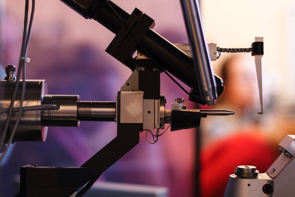
X-ray crystallography is a method of interpreting the structural arrangement of crystals at the atomic scale based on the generated diffraction pattern of scattered incident X-rays. The analytical power of X-ray crystallography extends from bulk scale crystals, such as minerals and metals, to investigation of bonding between small organic molecules and the three-dimensional structure of biomolecules such as proteins.

Image Credit: Gregory A. Pozhvanov/Shutterstock.com
This article will discuss the function and applications of X-ray crystallography and some of its specialized variations.
The Art and Science of Crystallography
A crystal is a solid material whose constituent components, be they atoms, molecules, ions, or other repeating monomeric units, are arranged in a regularly repeating structure, with orientation and monomer-monomer bonding determined by the electromagnetic, chemical, and physical properties of the monomer.
True crystals are constructed purely from one or more repeating monomers and maintain the same arrangement in all directions, while a collection of these crystals with grain boundaries and two-dimensional defects in a crystal structure are technically termed polycrystals. A single slow flake, for example, is a crystal, while a typical ice cube is likely to be a polycrystal.
When an X-ray strikes an atomic nucleus directly, it undergoes elastic scattering, meaning that its energy is conserved, but its direction of propagation is changed. X-rays are smaller than the gaps between atoms in a crystal and thus are able to penetrate multiple layers and potentially engage in scattering interactions with deeper layers of atoms in the same manner as surface atoms, scattering in the same direction of propagation, as determined by the angle of incidence.
Owing to the regular arrangement of atoms in a crystal, X-ray scattering patterns are produced, known as a diffraction pattern, made up of intense spots where X-rays come together in constructive interference and spots lacking intensity owing to destructive interference. The diffraction pattern can, therefore, be used to infer the spacing between atoms within the crystal structure.
From Crystals to Molecules
X-ray crystallography has been used extensively since the 1920s in determining the arrangement of atoms in crystal structures, such as minerals and metals. Also, as early as the 1920s, X-ray crystallography had been applied to the determination of molecular structures with lengthy repeating units and relatively simple atomic arrangements, such as long-chain fatty acids.
In order to engage in X-ray crystallography of flexible molecules such as this, they must first be purified extensively and brought as close as possible to the point of supersaturation by concentration and precipitation of the molecule from solution. This is typically achieved by vapor diffusion, wherein a small drop containing the molecule of interest is placed in a drying chamber that steadily evaporates away the solvent.
Using these methods, the first protein structure was elucidated by X-ray crystallography in the 1950s, that of sperm whale myoglobin. Since then, the technique has become the leading method in the determination of protein structure and is used extensively throughout the life sciences and pharmaceutical industries in investigation of small molecule and biomolecular interactions.
X-ray Crystallography Techniques
The type of X-ray crystallography described so far is termed single-crystal X-ray crystallography, wherein, ideally, the spatial arrangement of atoms in the purest and most regularly arranged crystals can be described with extreme resolution, even able to detect the minute vibrations of individual atoms. More complex or much smaller crystal structures than bulk materials, such as biomolecules or individual organic molecules, generate a confusing or weak diffraction pattern that may not be easily interpreted. Oriented chains of biological molecules such as fatty acids, for example, may appear as “bands” of diffraction patterns that are indicative of macromolecular structure but not as useful in the precise determination of atomic distances.
Powder X-ray diffraction is operationally similar to single-crystal X-ray crystallography, the difference being the state of the sample, which is powdered and, therefore, contains crystal grains of every possible orientation relative to the incident X-rays.
Within this mixture, a significant proportion are coincidentally in the correct orientation to diffract X-rays towards the detector at any given moment, but due to the random mixture of angles between layers of atoms present within the sample, a ring of diffraction spots is generated instead of a single spot. A diffractogram is generated by constructive and destructive interference in powder X-ray diffraction, and mathematical Fourier-transform procedures are utilized to interpret the signal.
Various additional analytical methods have been developed based on the principles of X-ray crystallography, such as neutron diffraction. Relatively low-energy X-rays used in ordinary X-ray crystallography are only able to penetrate a few upper layers of the crystal, while higher energy radiation generated in synchrotrons can penetrate further. Neutrons are able to penetrate much more deeply into the material owing to their innate lack of interaction with materials, which they largely pass through.
Beyond 3D: Advancements and Challenges in X-ray Crystallography
X-ray crystallography is faced with a number of challenges outside of the characterization of the upper layers of pure crystals with well-defined, ordered structures. Innovations surrounding the refinement of X-ray generators and detectors and the subsequent mathematical treatment of the generated interferogram have further pushed the abilities of technique into the regular characterization of complex molecules such as proteins and their interaction with drug compounds and larger biological systems.
Three-dimensional structures such as nanoparticles and microcrystals, as well as weakly diffracting crystals, are still difficult to analyze by single-crystal X-ray crystallography, though this can be compensated somewhat by serial analysis. Serial X-ray crystallography can be used to gather data at higher resolution and also over time, allowing analysis of moving biological systems, such as changes in protein conformation in response to external stimuli.
Further Reading