Assays are important analytical processes that are involved in many aspects of biological laboratory work. They are a way of evaluating the contents of samples and actions of various molecules and are important for diagnoses, disease understanding, drug discovery, and therapeutic development.

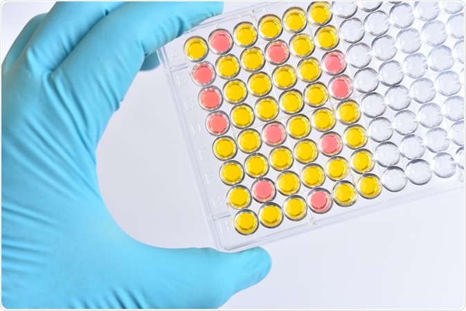
Assay. Image Credit: Jarun Ontakrai/Shutterstock.com
There are many different types of assay, each having distinct uses, benefits, and drawbacks. Assays can be divided into three main categories based on the type of sample used – ligand-binding assays that measure binding between a ligand and a receptor, immunoassays that detect antibody-antigen binding, and bioassays that measure biological activity in response to certain stimuli.
Assay outputs
Assays produce an array of outputs depending on the technique used. When required for diagnostic purposes, a qualitative output is most useful to determine if a certain disease marker is present or not. Quantitative outputs are also crucial for measuring the severity of diseases, the efficacy of treatments, or other continuous scale measurements, such as mineral levels in soil.
Some assay techniques can provide definitive quantity readings, while others require the generation of a standard curve to compare unknowns to. Functional assays are used to assess the function of substances, such as the potency of competitive drugs.
Different assay techniques
While the specifics of the process vary based on the type of assay technique being used, all in vitro assays follow a similar outline. First, a test sample must be collected and prepared. In many cases, the sample will be purified by a process such as filtration, ultracentrifugation, or selective binding to remove any substances that are not the test sample and avoid background interference.
Some simpler assays, especially those that are purely qualitative, may not require this step, particularly if the substance being tested for is present in high quantities in a sample. Once the sample is prepared, the test process itself begins, with the sample being introduced to the detection system, be that antibody testing in an enzyme-linked immunosorbent assay (ELISA), or immobilized receptors for a ligand-binding assay for example.
To reduce the possibility of false-positive results, the bound sample will be washed throughout the detection process to ensure that any unbound debris is removed. The signal will then be detected and measured, using an appropriate method for the technique used, such as colorimetry, radioisotope detection, or electrical detection. For this detection to occur, results frequently have to be amplified, often by binding of a secondary signal molecule.
Bioassay protocols will be different from other assay techniques due to the nature of the samples and tests being carried out. They may involve tissue sample histology, measurements of cell culture growth, or measurements of electrical or chemical output of tissues following stimulation.
Ligand-binding assays
A ligand-binding assay is a technique used to measure the binding ability of ligands to receptors. They are useful for assessing signaling activity in cell pathways and for predicting the affinity of potential new drugs. Most ligand-binding assays require the use of a radio-labeled ligand for quantitative detection, though ligand-binding assays can also use non-radioactive labeling.
Radio-label assays are frequently used to measure affinity by displacement. The ligand of interest, which is unlabelled, will be introduced to the receptor and any unbound ligand then removed. The radio-labeled ligand is then added, the assay is washed again and radioactivity can be measured. In this kind of affinity assay, an increased level of radioactivity measured indicates that the ligand of interest has a lower binding affinity to the receptor used than the radio-labeled ligand.
Radio-labeled ligand-binding assays can be used without signal amplification, reducing costs, though they require extensive washing and care with handling radioactive substances, so can be considered less desirable. Non-radioactive alternatives include using a fluorescent label for ligand-binding detection or using an enzyme-bound ligand and a substrate reaction, like those used in ELISAs.

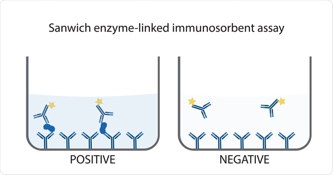
Sandwich enzyme-linked immunosorbent assay. Image Credit: Fluke Cha/Shutterstock.com
Immunoassays
Immunoassays are any assays that use antibodies or antibody fragments. One of the most used types of immunoassay is an ELISA. There are four main types of ELISA – direct, indirect, sandwich, and competitive – each of which has a slightly different function. The overarching principle of ELISA techniques is the use of an enzyme-conjugated antibody or antigen to measure binding to an immobilized antigen or antibody, respectively.
Binding levels can be assessed using a color-changing redox reaction involving a substrate that is added to the test being acted upon by the conjugation enzyme, which is commonly horseradish peroxidase. The reaction is then detected using a colorimeter and can be compared to a standard curve of known quantities.
Other immunoassays use markers that are not enzymes to show antigen-antibody binding. These markers can include fluorescent molecules and radiolabels, like those used in ligand-binding assays. Both ELISAs and other immunoassays are some of the most commonly used diagnostic assays, as a binary qualitative result, i.e. the presence or not of a disease-related antigen, is quick and relatively cheap to achieve.
Bioassays
Bioassay techniques are far more varied than other assays and range from in vivo experiments to assays which only require cell cultures. In vivo bioassay techniques are among the most prevalent assay techniques used in biological research and involve a wide variety of model organisms that each have different benefits.
These models organisms include mice, useful models because of their genetic similarity to humans, complex nervous system and relatively quick life cycle; zebrafish, which are ideal for studying embryonic development because their embryos are transparent; and fruit flies, due mainly to their rapid life cycles. Not all model organisms are animals, though, and microorganisms such as yeast and Escherichia coli are also used for bioassays.
In addition to in vivo studies, bioassays also include ex vivo studies, using tissue or organ samples, as well as in vitro experiments using laboratory-grown cell or tissue cultures. One of the most recent developments in bioassay techniques is that of wound healing simulation. This involves the growth of a tissue that can then be cut in a simulation of a wound and the repair mechanisms and rates can be studied.
References
- Cappiello, F., Casciaro, B. and Mangoni, M. L. (2018) ‘A novel in vitro wound-healing assay to evaluate cell migration.’ Journal of Visualized Experiments.
- De Jong, L. A. A., Uges, D. R. A., Franke, J. P. and Bischoff, R. (2005) ‘Receptor-ligand binding assays: Technologies and applications.’ Journal of Chromatography B: Analytical Technologies in the Biomedical and Life Sciences.
- Mayer, A. P. and Hottenstein, C. S. (2016) ‘Ligand-Binding Assay Development: What Do You Want to Measure Versus What You Are Measuring?’ AAPS Journal.
- Tang, B., Wang, Y., Zhu, J. and Zhao, W. (2015) ‘Web resources for model organism studies.’ Genomics, Proteomics, and Bioinformatics.
Further Reading