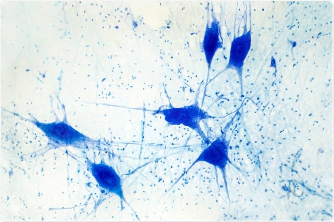
Fluorescence microscopy is a powerful tool used to investigate proteins of interest while preserving the integrity of the cell architecture. The advent of different fluorophores facilitates multiplexing, providing opportunities to conduct visual investigations such as co-localization. Preparing cells for immunofluorescence is somewhat empirical; it depends on the protein being investigated and the cell line/type being used. Each step requires consideration prior to investigation.

Image Credit: Kateryna Kon/Shutterstock.com
Sample preparation
Sample preparation depends on cell type, the protein(s) of interest (whether it is transfected or endogenous), and its subcellular location. Cell lines need to be treated differently if they are suspended or adherent. Critically, the samples must not be allowed to dry out or exposed to light. Humidified containers can be helpful in this regard.
Adherent cell lines behave differently in terms of replication, morphology, and adhesion. Therefore, these factors need to be considered before commencement. Cells are typically seeded at 50-60% confluency 12-18 hours prior to experimentation.
Coverslips or glass-bottomed cell culture dishes can be utilized; these can be pre-treated to promote adhesion. These include poly-Lysine, gelatin, Matrigel®, etc. Suspension cells need to be centrifuged and then allowed to adhere to coverslips prior to experimentation. Cell toxicity and cell death should also be considered before preparation, as they introduce artifacts.
Microscopy considerations
Prior to sample preparation, it is essential to investigate the lasers and filters associated with the microscope. Different lasers excite different fluorophores with a defined spectrum. Multiplexing is useful as it enables the labeling of multiple proteins of interest, but spectral overlap needs to be considered. Photo-bleaching and phototoxicity also must be considered as they can damage the sample.
Buffers
An important consideration before experimentation is the type of buffer used. Typically, it is slightly basic (pH 7.0-7.4), phosphate-based (e.g., Phosphate Buffer Saline), but specialist buffers can be used such as PEM, MOPS, TES, HEPES, and PIPES.
This will be used for washing but also for adulteration of the antibodies and other agents, so it is critical to investigate this. This is particularly important for washing steps. These occur after primary and secondary antibody incubation to prevent background; otherwise, this can introduce artifacts into the sample.
Fixation
The purpose of fixation is to generate a snapshot of the sample. This is particularly important for cell architecture or localization studies. It is, however, not useful for dynamic studies. Fixatives tend to be crosslinking reagents or organic solvents.
The type of fixative depends on the sample: many can affect the strength of the signal. It is critical to consider the proteins of interest as this can predicate fixation agents.
Denaturing fixatives such as methanol and acetone cannot be used with GFP-tagged proteins as they extinguish the signal. Due to the denaturing aspect of these, they contribute to the exposure of epitope regions but also impair the 3-Dimensional protein structure. Paraformaldehyde is typically used in immunofluorescence (at 4%), but it dissolves lipids in the membrane. Glutaraldehyde is typically used for electron microscopy.
Other factors to consider are duration and temperature. In standard protocols, this is performed for 20 minutes at room temperature. These parameters can be modulated, especially in very mobile proteins such as the cytoskeleton.
Permeabilization
Due to the nature of the plasma membrane, it is desirable to use a permeabilizing agent to perforate the membrane and facilitate the ingress of antibody molecules. These tend to be detergents that are amphiphilic and thus facilitate interaction with both hydrophobic and hydrophilic moieties.
Non-ionic detergents include Triton X-100, Tween 20, and Nonidet P-40. These are typically utilized at 0.1%. Ionic detergents solubilize the membrane but also affect the 3-D structure. These include SDS, CHAPS, and deoxycholate. Saponins are also used as they preserve ultrastructural integrity. These are usually only used for a short duration at room temperature, but this is dependent on the sample.
Blocking
Blocking is used to inhibit binding to non-specific sites. Reagents include Bovine Serum Albumin, non-fat milk, or serum. Blocking solutions are incubated at room temperature for 30-60 minutes. It is recommended that blocking proteins do not originate from the same species as the primary antibody is raised.
Detection
Detection of the protein of interest can be achieved using direct or indirect detection. In direct immunofluorescence, the fluorophore is directly conjugated to the primary antibody. Indirect fluorescence involves a secondary antibody (with a fluorophore) that binds specifically to the primary antibody.
Once again, this decision needs to be undertaken prior to experimentation in consultation with the published literature. Indirect IF is more popular due to high sensitivity, signal amplification, and multiplexing. The primary antibody must be raised against an epitope in the target protein while the secondary antibody is raised in a different species to the primary. Poly- or monoclonal antibodies can be applied.
Polyclonal antibodies bind to different epitopes in the protein of interest, while the mono-clonal bind to one epitope specifically. This can be critical for signal detection and specificity. Antibodies tend to be used at room temperature for one to two hours at concentrations between 1:100 and 1:1000. Overnight incubation at 4 °C may be feasible (Im et al., 2019).
Final considerations
Stains that intercalate DNA such as DAPI or Hoescht can be used to visualize the nucleus. It is important to note their excitation spectrum in multiplexing. Mounting the samples for microscopy involves an anti-fade mountant.
Please refer to the manufacturer’s instructions as some warrant sealing. Samples are then typically stored at 4°C, in the dark.
Controls
Controls can be incorporated into the protocol to mitigate potential artifacts. A sample that has been prepared but no antibodies have been applied provides information about autofluorescence.
A sample should be incubated with a secondary antibody only to determine non-specific binding. This can then be used to determine the threshold parameters for the microscope. If multiple fluorophores are being utilized individual fluorophores can be applied singly to determine crosstalk between spectra.
Sources:
- Burry, R. W. (2011) ‘Controls for immunocytochemistry: an update’, The journal of histochemistry and cytochemistry : official journal of the Histochemistry Society, 59(1), pp. 6–12. doi: 10.1369/jhc.2010.956920.
- Donaldson, J. G. (2015) ‘Immunofluorescence Staining’, Current Protocols in Cell Biology, 69(1), pp. 4.3.1-4.3.7. doi: https://doi.org/10.1002/0471143030.cb0403s69.
- Im, K. et al. (2019) ‘An Introduction to Performing Immunofluorescence Staining’, Methods in molecular biology (Clifton, N.J.), 1897, pp. 299–311. doi: 10.1007/978-1-4939-8935-5_26.
- Jamur, M. C. and Oliver, C. (2010) ‘Permeabilization of cell membranes.’, Methods in molecular biology (Clifton, N.J.), 588, pp. 63–66. doi: 10.1007/978-1-59745-324-0_9.
- Kudalkar, E. M. et al. (2016) ‘Coverslip Cleaning and Functionalization for Total Internal Reflection Fluorescence Microscopy’, Cold Spring Harbor protocols, 2016(5), p. pdb.prot085548-pdb.prot085548. doi: 10.1101/pdb.prot085548.
- Pereira, P. M. et al. (2019) ‘Fix Your Membrane Receptor Imaging: Actin Cytoskeleton and CD4 Membrane Organization Disruption by Chemical Fixation’, Frontiers in Immunology, p. 675. Available at: https://www.frontiersin.org/article/10.3389/fimmu.2019.00675.
Further Reading
Last Updated: Oct 5, 2021