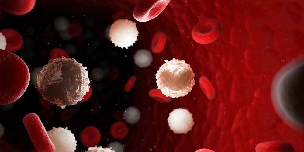
In this article, AZoLifeSciences explores how flow cytometry can be applied to the diagnosis of leukemia.

Image Credit: SciePro/Shutterstock.com
What is Leukemia?
Leukemia, which is the 11th most common type of cancer to affect human beings across the world, is a malignancy of the bone marrow. Leukemia can be further categorized into several subtypes that fall into the category of either “acute” or “chronic” forms of leukemia.
Acute leukemias, for example, are derived from early progenitor cells in the bone marrow and neoplastic cells and have an immature blast-like appearance. Comparatively, chronic leukemias, while also derived from defective early cells, retain the capacity to differentiate into cells that resemble normal end-stage mature blood cells. Typically, the differentiation between acute and chronic leukemias is made based on the morphological assessment of the patients’ blood and bone marrow samples.
In addition to the acute and chronic classifications for leukemias, further subtypes can be determined based on the antigenic profile of the affected cell lineage and the cell's maturation or differentiation stage. This can therefore include B-lymphoid, T-lymphoid, or myeloid subtypes of both chronic and acute leukemia. For this sub-classification, immunohistochemistry or flow cytometry can be used to distinguish between these subtypes of leukemia.
How Flow Cytometry is Used for Leukemia Diagnosis
Flow cytometry allows researchers and clinicians to analyze and characterize blood cells according to the expression of specific surface antigens. To achieve this, various fluorochrome-conjugated antibodies are incorporated into a blood sample, each of which is designed to label a specific antigen of interest.
The labeled sample is then injected into the flow cytometer, wherein several lasers provide excitation of the fluorochromes, which subsequently allows the detectors of this system to quantify the emitted fluorescent signals. Thus, the investigator is able to identify specific lymphoid and myeloid populations present within a peripheral blood sample, as well as verify the expression of specific antigenic determinants, to arrive at a final diagnosis of the type of leukemia and its appropriate subclassification.
Diagnosing Acute Leukemias with Flow Cytometry
As previously mentioned, acute leukemia refers to a malignancy of abnormal, immature white blood cells. Typically, acute leukemia is categorized as either acute myeloid leukemia (AML), which affects cells like monocytes and neutrophils that are of the myeloid lineage, as well as precursor lymphoid neoplasms. The second subtype of acute leukemia is acute lymphoblastic leukemia (ALL), which affects cells of the B- or T-lymphoid lineage.
Regardless of the subtype, the diagnosis of any acute leukemia must include information on the morphology of the blood and bone marrow cells, as well as immunophenotyping, cytogenetics, and molecular genetics. Importantly, immunophenotyping that is achieved through the use of flow cytometry is an essential aspect of the diagnostic workup for acute leukemias.
To date, standard flow cytometry can provide immunophenotypic information on cells based on antigen expression that is detected with fluorescent antibodies and offers the user the ability to assess multiple fluorescent parameters simultaneously. It should be noted that standard flow cytometry is limited in its ability to visualize cells directly and is, therefore, unable to distinguish the pattern and localization of antigenic expression on cells.
Imaging flow cytometry has emerged as an alternative diagnostic technique that offers superior information as compared to when standard flow cytometry is used in the diagnosis of acute leukemias.
For example, imaging flow cytometry is capable of visualizing cell morphology, analyzing differences in cell size and shape, and determining the cellular location and pattern of antigen expression. Each of these parameters, in addition to those that are accomplished when standard flow cytometry is used alone, are assessed simultaneously.
Diagnosing Chronic Leukemias with Flow Cytometry
The two major types of chronic leukemia include chronic lymphocytic leukemia (CLL) and chronic myeloid leukemia (CML). Despite the fact that CLL is the most common type of adult leukemia in the Western World, its treatment is highly complex and often determined based on the presence of specific molecular markers.
The diagnosis for CLL can only be confirmed when at least 5 x 10⁹ L/B lymphocytes have been detected in the peripheral blood, in addition to identifying a clonal B-cell population. Flow cytometry is a common technique that is incorporated into the diagnostic methods for CLL. In fact, the most common flow cytometry-based prognostic markers used for CLL diagnosis include CD38 and ZAP70, both of which are proteins involved in B-cell receptor signaling, as well as the presence of the surface integrin CD49d.
Flow cytometry is often relied upon for the diagnosis of CML as a result of its ability to capture, label, and detect the BCR-ABL1 fusion protein. However, despite the utility of flow cytometry in diagnosing many hematologic malignancies, there is no explicit role that has been designated for flow cytometry by the World Health Organization (WHO) in the diagnosis of one subtype of CML known as chronic myelomonocytic leukemia (CMML). Recent efforts have been made to include monocyte subset quantification in clinical flow cytometry panels to assist in the diagnosis of CMML. One advantage to using flow cytometry for this purpose is that it is a much more simple system to operate as compared to genetic analyses.
References and Further Reading
Grimwade, L. F., Fuller, K. A., & Erber, W. N. (2017). Applications of imaging flow cytometry in the diagnostic assessment of acute leukaemia. Methods 112; pp. 39-45. https://www.sciencedirect.com/science/article/abs/pii/S1046202316301980
Scheuermann, R. H., Bui, J., Wang, H., & Qian, Y. (2017). Automated Analysis of Clinical Flow Cytometry Data: A Chronic Lymphocytic Leukemia Illustration. Clinics in Laboratory Medicine 37(4); pp. 931-944. https://www.sciencedirect.com/science/article/abs/pii/S0272271217300732
Strati, P., Jain, N., & O-Brien, S. (2018). Chronic Lymphocytic Leukemia: Diagnosis and Treatment. Mayo Clinic Proceedings 93(5); pp. 651-664. https://www.sciencedirect.com/science/article/abs/pii/S002561961830154X
Hudson, C. A., Burack, W. R., & Bennett, J. M. (2018). Emerging utility of flow cytometry in the diagnosis of chronic myelomonocytic leukemia. Leukemia Research 73; pp. 12-15. https://www.sciencedirect.com/science/article/abs/pii/S0145212618301930
Radich, J., Yeung, C., & Wu, D. (2019). New approaches to molecular monitoring in CML (and other diseases) blood 134(19); pp. 1578-1584. https://www.sciencedirect.com/science/article/pii/S0006497120739923