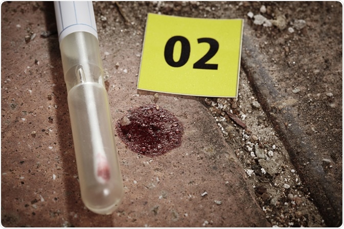
The ability to differentiate between different cell populations present within any type of biological evidence obtained from a crime scene is crucial to any investigation.

Image Credit: Couperfield/Shutterstock.com
Several different analytical techniques have been used for this purpose, some of which include histological staining, mRNA expression, DNA methylation analysis, and fluorescence-activated cell sorting.
The need for cell differentiation in forensics
Consider a sexual assault case involving a woman who has accused the defendant of using a bottle to sexually violate the victim.
During the collection of evidence from the crime scene, forensic scientists obtain the weapon of interest, which in this case would be the water bottle, and confirm that the DNA profile obtained from the trace evidence found on the bottle matches that of the victim.
To defend their client, the defense may assert that the DNA found on the bottle arose due to the victim drinking from the bottle, whereas the prosecution may instead allege that the DNA was vaginal in its origin.
These types of criminal cases, therefore, require thorough cell identification methods capable of differentiating the specific cellular origin.
Histological techniques
The three most relevant cell types that are used as forensic evidence include skin, buccal and vaginal epithelial cells, all three of which are classified as stratified squamous epithelium.
Although skin epithelial cells are keratinized and lack nuclei, buccal and vaginal cells are practically indistinguishable from one another, as both of these cell types contain nuclei. Histology stains are frequently used in a forensic laboratory; therefore, this method was one of the first applied to this challenging situation.
Several different histological techniques have been employed to differentiate these three cell types from one another. One of the earliest attempts to do this was achieved by Lugol’s iodine, which was developed during the 1970s.
Positive staining for Lugol’s iodine represented the presence of glycogen granules, which ultimately indicated vaginal cells. Despite its usefulness, this method has been found to also confirm the presence of glycogenated squamous cells within the oral mucosa, male urinary tract, and the penis.
In a 2008 study, eleven different histological stains, some of which included Dane’s Csaba’s and Ayoub-Shklar stains, were evaluated for their effectiveness in differentiating between these three stratified squamous epithelial tissue samples.
Of all of the stains tested in this study, Dane’s stain, which primarily stains for prekeratin, keratin and mucin proteins in tissue samples, was found to provide different staining patterns for skin, buccal and vaginal smeared cells.
mRNA sequencing
Messenger RNA (mRNA) is a single-stranded RNA molecule that is complementary to a single DNA strand in a given gene. During translation, mRNA is decoded by ribosomes to produce a specific amino acid chain, which will ultimately contribute to the formation of an active protein. The significance of studying mRNA expression is attributed to its unique ability to predict protein expression. One of the most widely used methods for mRNA expression quantitation is a real-time reverse transcriptase-polymerase chain reaction (RT-PCR).
Within the field of forensic science, RT-PCR has been widely used to assess the expression of various mRNA markers in forensically relevant body fluids like saliva, blood, semen, vaginal secretions, and menstrual blood. Table 1 provides a brief overview of tissue-specific genes that have been identified in these critical human tissues.
|
Human Tissue
|
Gene
|
|
Blood
|
Porphobilinogen deaminase (PBGD)
Beta-spectrin (SPTB)
|
|
Saliva
|
Statherin (STATH)
Histastin 3 (HTN3)
|
|
Semen
|
Protamin 1 (PRM1)
Protamin 2 (PRM2)
|
|
Vaginal secretions
|
Human beta-defensin 1 (HBD-1)
Mucin 4 (MUC4)
|
|
Menstrual blood
|
Matrix metalloproteinase 7 (MMP-7)
Matrix metalloproteinase 11 (MMP-11)
|
Fluorescence-activated cell sorting
Fluorescence-activated cell sorting (FACS) is widely used in the medical setting for diagnostic and research purposes. Through the assistance of fluorescent probes, FACS allows users to sort cells based on selected characteristics of interest.
Despite its usefulness, FACS has rarely been used for the analysis of forensic samples; however, several efforts have been made to make FACS a mainstream method.
Some of the earliest applications of FACS analysis in forensic science included its use to separate mixed sperm and vaginal epithelial cell populations. Also, to begin a costly technique at the time, this preliminary attempt only utilized markers that were specific to sperm cells, thereby limiting the validity of this method.
As cell sorting machines have undergone considerable technological advancements over the past several decades, cell sorting has become more sensitive to a point where single-cells can be isolated and identified. In a 2015 study, a group of Australian researchers utilized FACS for the separation of cellular mixtures before DNA extraction.
To this end, 14 different ratios of blood and saliva mixtures were analyzed by FACS. To, identify saliva-based epithelial cells, anti-CD227 was used, whereas blood-derived leukocytes were targeted with an anti-CD45 probe.
This work not only demonstrated the usefulness of FACS for separating fresh blood and saliva mixtures but also proved useful in improving the number of detectable alleles from targeted cell types during subsequent DNA extraction and analysis.
The value of this concept has been proven; however, more research is essential before FACS could be applied to actual forensic samples.
Sources
- French, C. E. V., Jensen, C. G., Vintiner, S. K., Elliot, D. A., & McGlashan, S. R. (2008). A novel histological technique for distinguishing between epithelial cells in forensic casework. Forensic Science International 178(1); 1-6. DOI: 10.1016/j.forsciint.2008.01.010.
- Haas, C., Klesser, B., Kratzer, A., & Bar, W. (2008). mRNA profiling for body fluid identification. Forensic Science International: Genetics Supplement Series 1(1); 37-38. DOI: 10.1016/j.fsigss.2007.10.064.
- Brocato, E. R., Philpott, M. K., Connon, C. C., & Ehrhardt, C. J. (2018). Rapid differentiation of epithelial cell types in aged biological samples using autofluorescence and morphological signatures. PLOS One. DOI: 10.1371/journal.pone.0197701.
- Verdon, T. J., Mitchell, R. J., Chen, W., Xiao, K., van Oorschot, R. A. H. (2015). FACS separation of non-compromised forensically relevant biological mixtures. Forensic Science International: Genetics 14; 194-200. DOI: 10.1016/j.fsigen.2014.10.019.
Further Reading