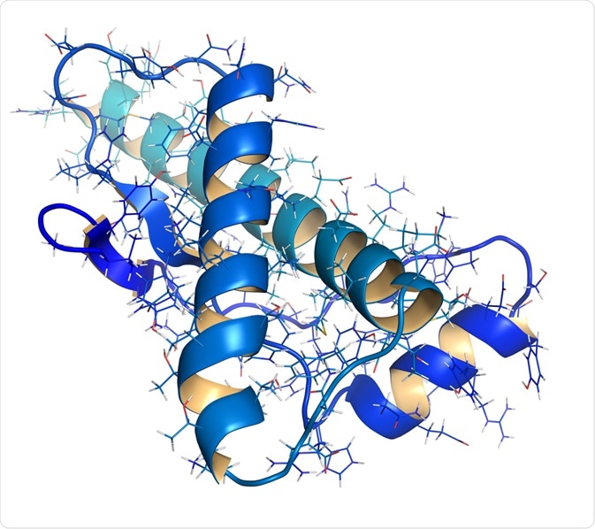
This December, a team of Australian researchers published findings from their new study in the scientific journal Angewandte Chemie International Edition.

Image Credit: StudioMolekuul/Shutterstock.com
In their paper, they describe how they have established a new method of viewing the folding state of proteins in vivo, equipping scientists with a tool to measure cellular health.
The team developed a molecular probe that can feedback data on unfolded proteins by mapping the subcellular polarity of the unfolded proteome environment. Using a fluorescent signal, the technique can quantify the unfolding of proteins in the cell.
This innovative tool is likely to give insights into the signaling pathways of diseases such as cancer and neurodegeneration.
Understanding protein misfolding may impact future disease treatment
As part of their normal life cycle, proteins undergo conformational changes that lead to misfolding, for example during the processes of synthesis and degradation.
However, changes in protein conformation do not always go smoothly, and abnormalities in this process have been linked with various diseases. Aggregates of misfolded proteins are characteristic of neurodegenerative disorders, such as Alzheimer’s and Parkinson’s disease.
Recent studies have highlighted how the process of protein folding itself is closely linked to cancer and that the molecules involved in such a process could potentially act as therapeutic targets for new cancer treatments. Recent research has demonstrated that to proliferate rapidly, cancer cells utilize the same chaperone molecules that are responsible for correct protein folding.
Scientists have suggested that targeting of specific chaperones as HSPs may stop cancer without harming healthy cells since cancer cells rely much more heavily on these molecules.
With protein folding and misfolding is implicated in numerous diseases that impact on a vast number of people, understanding the mechanisms regulating these processes becomes essential to enable the development of new therapies.
Currently, research has uncovered that protein folding mechanisms become imbalanced in stressed cells, resulting in errors in the folding process.
This creates misfolded proteins, which become trapped in the endoplasmic reticulum or are degraded prematurely, resulting in protein aggregation and debris, which are the hallmarks of neurodegenerative disease and cancer.
Earlier studies have determined that subcellular polarity is a driving factor of protein misfolding and aggregation. This inspired the work of the team at the University of Melbourne, Australia, who aimed to establish a method for monitoring protein aggregation based on measuring misfolded proteins with fluorescent probes.
They recognized that the impact of innovation in this field could be significant for enabling better treatment options for various cancers and neurodegenerative diseases.
The development of a two-modal fluorogenic probe
The team designed a two-modal fluorogenic probe capable of monitoring protein aggregation in unprecedented detail. When operating in the first mode, the probe senses misfolded proteins by binding to binds to the exposed cysteine residues on the surface of misfolded proteins.
These residues are usually encased deep within correctly folded proteins where they form stable bridges. This binding activity triggers fluorescence, which can then be measured.
When functioning in the second mode, the probe measures the polarity of the subcellular environment. It is known that polar environments are a result of unbalanced electronic distribution.
The probe assesses polarity by accessing data on the dielectric constant. To achieve this, the researchers added a "push-pull" chemical group to the fluorogenic probe.
They observed that the fluorogenic probe emitted its fluorescence signal with a shift in color in response to polar solutions with a high dielectric constant.
Probe visualizes misfolding in unprecedented detail
The probe developed by the researchers was named NTPAN-MI and was tested on human cells which were put under stress by introducing drugs known to interfere with the processes of protein synthesis and folding. Under test conditions, the team saw that untreated cells emitted normal fluorescence, but in response to unfolded or misfolded proteins accumulated in cells treated with toxicants or infected with a virus, a bright fluorescence was emitted.
The team also observed color shifts that signaled the polarity of the environment. Impressively, the team reported that for the very first time they were able to visualize the "unfolded protein load" in the nucleus. Previous methods could only measure unfolded or misfolded proteins in the cytoplasm.
What’s next?
The establishment of a reliable and effective method for measuring unfolding and the polarity of the protein environment will be significant in advancing our knowledge of the process of protein folding, and how this process goes wrong.
This will likely be instrumental in developing new therapies for diseases characterized by protein misfolding, such as cancer and neurodegenerative diseases. Before this can happen, more research is needed to further develop this method and use it in other applications.
Sources:
- Owyong, T., Subedi, P., Deng, J., Hinde, E., Paxman, J., White, J., Chen, W., Heras, B., Wong, W. and Hong, Y. (2020). A Molecular Chameleon for Mapping Subcellular Polarity in an Unfolded Proteome Environment. Angewandte Chemie International Edition. https://onlinelibrary.wiley.com/doi/abs/10.1002/anie.201914263
- Sweeney, P., Park, H., Baumann, M., Dunlop, J., Frydman, J., Kopito, R., McCampbell, A., Leblanc, G., Venkateswaran, A., Nurmi, A. and Hodgson, R. (2017). Protein misfolding in neurodegenerative diseases: implications and strategies. Translational Neurodegeneration, 6(1). https://www.ncbi.nlm.nih.gov/pmc/articles/PMC5348787/#!po=52.1277
- Van Drie, J. (2011). Protein folding, protein homeostasis, and cancer. Chinese Journal of Cancer, 30(2), pp.124-137. https://www.ncbi.nlm.nih.gov/pmc/articles/PMC4013342/#!po=70.6897
Further Reading