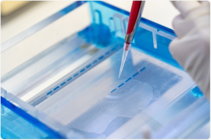
Electrophoresis is a technique in which an electric current is applied, driving the separation of biological molecules, including DNA/RNA and proteins in a gel-based process. The separation of molecules is based on differences in velocity. This relates to molecule mobility and field strength. This mobility is governed by particle size, shape, and charge which remain constant during electrophoresis.

Image Credit: Choksawatdikorn/Shutterstock.com
In the case of nucleic acids, they possess an intrinsic negative charge which promotes their migration in the direction of the positive electrode (anode). Several factors can affect the separation of molecules. The size and conformation of the DNA affect its migration. Larger fragments migrate slower as they experience greater resistance forces.
Supercoiled DNA migrates faster than relaxed DNA. The gel concentration also affects the process: higher concentrations generate a tighter matrix with smaller pore sizes, inhibiting the migration of larger fragments. Agarose is the most common gel used and is suitable for the resolution of fragments between 10 kilobases and 0.1kb (concentrations between 0.5% and 3% w/v in appropriately buffered solutions such as TAE).
For the resolution of smaller fragments PAGE gels are recommended. The strength of the current applied also affects the resolution. Higher voltages promote faster migration; they can also generate heat which affects resolution. The samples are labeled with a DNA-intercalating agent which visualizes the DNA under UV fluorescence, such as ethidium bromide or newer commercially available alternatives. Ethidium bromide is toxic and can affect the labeling of nucleic acids.
Adaptations of Gel Electrophoresis
Several adaptations of traditional gel electrophoresis have been generated, often for specific niche applications. Pulsed-field gel electrophoresis is commonly used to identify specific bacterial species. It is used for microbial infection control and food safety surveillance. It involves subjecting the digested genomic material of bacterial colonies to a series of alternating electric fields.
The rearrangement of the cathodes allows the mega-base fragments to migrate towards the anode in a size-dependent manner. This generates a unique profile of the chromosomal genomic material, allowing for the detection of specific bacterial strains.
Electrophoretic mobility shift assay is another important application of electrophoresis. A subset of proteins interacts with nucleic acids through specific moieties. To determine this, EMSA is often utilized. The protein of interest is titrated in increasing concentrations with nucleic acid. An electric pulse is then applied.
If the nucleic acid binds to the protein its mass-to-charge ratio will change resulting in a shift in its migration: it will now migrate slower. This contrasts with the control “free” nucleic acid that will migrate as normal.
What is Gel Electrophoresis? | miniPCR bio™
Electrophoresis in Research
Electrophoresis is arguably the most important in pre-clinical research due to its associated low costs and ease of use. Capillary electrophoresis and Next Generation Sequencing are becoming more ubiquitous in clinical diagnostics and forensics, but in research, these are still inaccessible for many research groups. PCR and electrophoresis remain important confirmation tools, and for cloning.
The use of bacterial transformation to generate constructs of interest is a tool used in most research laboratories. Electrophoresis is used to confirm the amplification of a DNA fragment of interest. To facilitate the introduction of a DNA fragment into a vector of interest the plasmid and the fragment must have complementary ends. This is facilitated by restriction enzymes that cleave specific sequences of DNA.
DNA ligase then serves to join the complementary ends. This restriction digest can be used in a confirmatory manner to determine the fragment was correctly inserted into the plasmid and correctly transformed into the bacteria. Confirmatory electrophoresis is also important for CRISPR-Cas 9 gene editing and cloning.
Electrophoresis can be used to detect RNA contamination in genomic DNA. The integrity of total RNA can be detected using electrophoresis: RNA is composed of 28S and 18S rRNAs in a ratio of 2:1. The presence of smearing indicates RNA degradation. Many processes require fragmented genomic DNA such as NGS and Chromatin Immunoprecipitation (CHIP); electrophoresis allows for the detection of fragments of the appropriate size.
Forensics
Most modern forensics for the detection of DNA at crime scenes involves the use of DNA fingerprinting. This involves the detection of a series of STRs (short tandem repeats) which are unique to individuals.
Following the amplification of DNA fragments, samples are subjected to capillary electrophoresis. This adapted form of electrophoresis allows for improved efficiency, mitigated human error, and requires reduced sample volumes. This method uses the same premise as regular electrophoresis: samples are separated in a capillary tube using a matrix (such as linear polysaccharide, polydimethyl-acrylamide, hydroxy-ethyl-cellulose with Polyvinylpyrrolidone).
The fragments are detected by fluorescence. Improvements in microfluidics allow for the separation and subsequent detection of DNA fragments using four-color labeling fluorescence detection.
Sources:
- Durney, B. C., Crihfield, C. L. and Holland, L. A. (2015) ‘Capillary electrophoresis applied to DNA: determining and harnessing sequence and structure to advance bioanalyses (2009-2014)’, Analytical and bioanalytical chemistry. 2015/05/03, 407(23), pp. 6923–6938. doi: 10.1007/s00216-015-8703-5.
- Hellman, L. M., and Fried, M. G. (2007) ‘Electrophoretic mobility shift assay (EMSA) for detecting protein-nucleic acid interactions’, Nature Protocols, 2(8), pp. 1849–1861. doi: 10.1038/nprot.2007.249.
- Lee, P. Y. et al. (2012) ‘Agarose gel electrophoresis for the separation of DNA fragments’, Journal of visualized experiments : JoVE, (62), p. 3923. doi: 10.3791/3923.
- Sharma-Kuinkel, B. K., Rude, T. H. and Fowler Jr, V. G. (2016) ‘Pulse Field Gel Electrophoresis’, Methods in molecular biology (Clifton, N.J.), 1373, pp. 117–130. doi: 10.1007/7651_2014_191.
Further Reading
Last Updated: Sep 9, 2021