Stabilizing emulsions of hydrophobic and hydrophilic liquids is useful for drug delivery as much as food texture control. A common method is to stabilize liquid-liquid interfaces with polymers of opposite charge, which form coacervate networks through electrostatic interactions while being adsorbed to the liquid-liquid interface.
In this case study, Swiss researchers developed a new method to analyze such interfaces during coacervate formation, by employing an interfacial shear rheology method while simultaneously injecting the coacervate forming polymers.

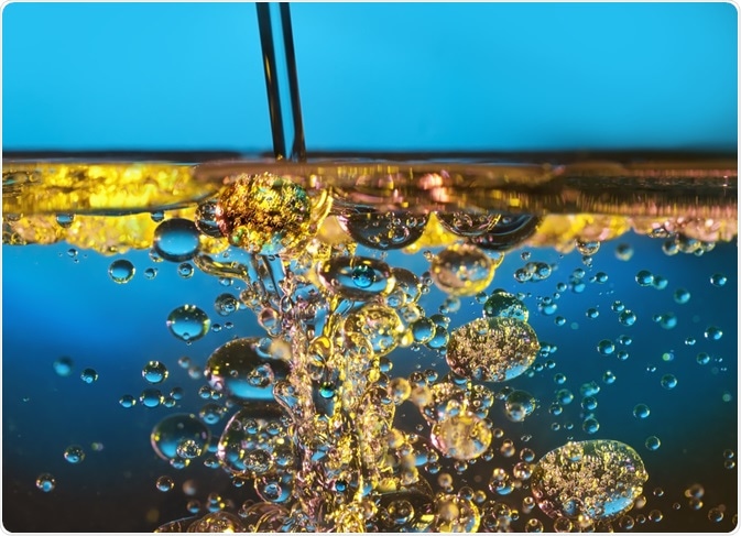
Image Credit: Mr.Lightman1975/Shutterstock.com
The texture of many foods is determined by controlled emulsification of hydrophobic and hydrophilic phases, with prominent examples being milk, yogurt, and butter. In such foods, a hydrophobic lipid phase, or droplet, is stabilized in an aqueous environment. However, different temperatures or shear force can irreversibly alter the phase separation.
Equally, many drug delivery formulations rely on controlled emulsification. For instance, a drug can be enclosed in a hydrophobic liquid, suspended as droplets in an aqueous environment, so that the drug becomes released only over longer periods.
To stabilize the liquid-liquid interfaces in emulsions against shear force or temperature-induced deformation, polymers can be bound to the interface. If different polymers are used, these can be applied as a mixed interfacial layer, or a multilayer, where different polymers are deposited layer-by-layer from the interface.
An example of such polymers is the combination of the protein β-lactoglobulin (β-lg) and the negatively charged sugar polymer pectin. β-lg is a major whey protein in cow milk that binds to lipids and stabilizes lipid phases in milk.
Previous research has demonstrated that it also forms interfacial layers at hydrophobic organic solvent interfaces with water. Pectin, often used with partial methoxylation, is a hydrophilic anionic polysaccharide that binds to interfacial β-lg layers, thus forming double-layer coacervates.
The stabilization of liquid-liquid interfaces by polymer coacervates can be measured as a reduction in surface tension and a stability gain against phase mixing. The stability of pre-formed coacervate layers can be well studied for instance in rheometers that measure the deformation of the interface when a certain force is applied. However, less is known about the actual formation period of the coacervates, since common rheometers are not equipped with injection modules.
Researchers from Zurich and Viligen (Switzerland), led by Professor Peter Fischer, developed a measurement setup for transient interfacial shear rheology, where tension can be applied to the liquid-liquid interface while simultaneously injecting coacervate forming compounds (“sub-phases”).
This allowed the scientists to determine the viscoelastic properties of the interfacial coacervate network during its formation, aiding the assessment of the optimal injection sequence and conditions.
Understanding the Formation of Interfacial Coacervates
The scientists used a liquid-liquid interface between water and the organic solvent n-dodecane as a model. To form an interfacial coacervate, β-lactoglobulin (β-lg) from whey protein and the anionic sugar polymer pectin (with methoxylations to control the number of anionic charges) were used.
β-lg and the methoxylated pectin were added with syringe pumps either as mixture, or subsequently in individual solutions, and rheology was measured.
Next, the investigators analyzed interfacial coacervate networks at the air-water interface both with shear rheology, and the final coacervate with neutron reflectivity to determine the coacervate layer thickness.
Layered Coacervates are Stronger and More Viscoelastic
At low pH, where β-lg carries a net positive charge and the pectin polymer a net negative charge, the scientists observed a favorable reduction of the liquid-liquid interface surface tension when β-lg was added initially and the pectin polymer subsequently.
The researchers also demonstrated that the addition of a pectin layer onto the interfacial β-lg layer did not change the surface tension further, however, strengthened the layer by increasing its viscoelastic properties. When both components were injected as a pre-mix, the interfacial coacervate formed slower and was less strong than in the case of subsequent addition of the coacervate formers β-lg and pectin.
From neutron reflectivity analysis at the air-water interface, the investigators concluded that the binding of pectin to an interfacial β-lg layer compressed the β-lg layer, in turn changing its viscoelastic properties.
The interfacial shear rheology setup employed here, where the coacervate forming polymers can be injected during the rheology measurement, thus enables to test different coacervate formers regarding their ability to stabilize liquid-liquid interfaces.
While this is useful for compound screening, the liquid-liquid interface in a rheometer is typically plane, not droplet-shaped as in emulsions. Thus, additional work will be required to test compounds in actual emulsion stabilization experiments.
Source
- Bertsch P et al. (2019). Transient measurement and structure analysis of protein-polysaccharide multilayers at fluid interfaces. Soft Matter. DOI: 10.1039/c9sm01112a.