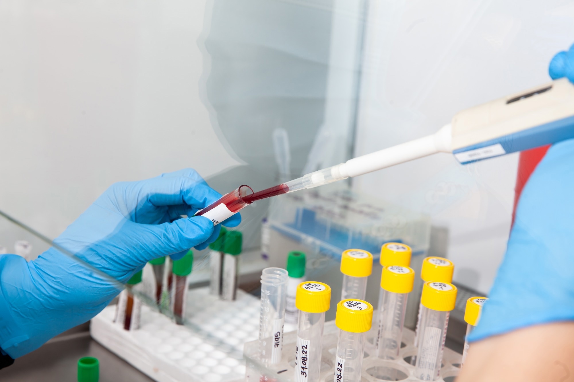
Fluorescence in situ hybridization (FISH) is a molecular cytogenetic technique that enables the detection and location of specific DNA sequences on chromosomes.1
It is also used to detect the presence of an entire chromosome in a cell. In contrast to other in situ hybridization (ISH) methods, this technique uses fluorescently labeled probes to detect specific DNA sequences.
 Image Credit: Anamaria Mejia/Shutterstock.com
History and Principles of the FISH Technique
FISH is a type of ISH technique developed by Joseph Gall and Mary Lou Pardue in the 1960s.2 This technique enabled the detection of DNA sequences in situ, i.e., within the chromosome.
Over the years, this technique was refined, increasing its sensitivity and versatility. Fluorescent labels were introduced in the 1970s and quickly gained popularity over radioactive isotopes because of their better stability, handling ease, and safety.
The main principle of FISH is associated with DNA hybridization. A FISH probe is a small DNA, RNA, or complementary DNA (cDNA)segment tagged with fluorophore molecules.3
A labeled probe and target DNA are denatured and mixed to allow complementary DNA sequences to anneal. The fluorescent probes bind to the target sequence due to high sequence complementarity. Fluorescence microscopy assesses fluorescence signals, which indicate location and specific gene expression in cells.4
Read More About Genetics and Genomics
Key Components of the FISH Technique
The three critical aspects of FISH are sample preparation, fluorescent probe, and fluorescent microscope. For a successful FISH experiment, it is crucial to prepare a slide with chromosomes in metaphase or interphase nuclei that are denatured at 70ºC.
Labeled probes are also denatured and introduced to the prepared slide. This slide is incubated at 37ºC for 5-15 hours for hybridization. After hybridization, this slide is visualized using Fluorescent microscopy.5
FISH uses different types of probes to detect target DNA sequences. Each probe is selected based on a specific application. For instance, Locus-Specific Probes (LPS) are used to locate chromosome genes.
This probe also identifies the number of genes expressed within a genome. Alphoid or centromeric repeat probes are another probe type that detects missing genetic material on a specific chromosome.6
Diversification of FISH Techniques
The original FISH protocol has been modified into various remarkable procedures.2 These modifications have tremendously improved this technique's sensitivity and specificity.
Besides tweaking the original FISH protocol, advancements in fluorescent microscopy and digital imaging have improved the understanding of chromatin and nucleic acids' physical and chemical properties. Some of the modified FISH technique types are presented in the table below.
Table 1: Different Types of Modified FISH Techniques and Applications2
| FISH |
Applications |
|
Centromere-FISH (ACM-FISH)
|
Detection of chromosomal abnormalities in sperm cells.
|
| Single Molecule RNA FISH (smFISH) |
Detection and quantification of the mRNA and other RNA molecules a thin layer of tissue sample |
| Cellular Compartment Analysis of Temporal (Cat) Activity by Fish (catFISH) |
Determine the interactions of neuronal populations associated with different behaviors. |
| armFISH |
Detection of chromosomal abnormalities in the p- and q-arms of all 24 human chromosomes. |
| Catalyzed Reporter Deposition-FISH (CARD-FISH) |
Detection, identification, and quantification of microorganisms involved in bioleaching processes. |
| Chromosome Orientation (CO)-FISH |
Assessing chromosomal translocations and inversions.
|
| Rainbow-FISH |
Simultaneous detection and quantification of up to seven different microbial groups in a microscopic field. |
| RNA-FISH |
Simultaneous detection, quantification, and localization of individual mRNA molecules either in the nucleus or cytoplasm at the cellular level in fixed samples. |
| e-FISH |
BLAST-based FISH simulation program can predict the outcome of hybridization experiments. |
| Flow-FISH |
Measurement of the telomeric signals from cells in suspension.
|
Advantages and Clinical Applications of FISH
FISH is a versatile technique used in wide-ranging clinical applications. The diverse applications of FISH have been attributed to its superior sensitivity, high specificity, and relatively low experiment time.
In medicine, FISH is used to diagnose and evaluate prognosis and treatment responses. Histiocytoid Sweet Syndrome is a dermatological disease that can be diagnosed using the FISH technique. For this diagnosis, it detects the presence of BCR/ABL gene fusion and chromosomal abnormalities in the cutaneous infiltrate of the initial biopsy specimen.7
The FISH technique is also used to diagnose pseudomosaicism from true mosaicism. Furthermore, it is used to diagnose Chronic Myeloid Leukemia, Chronic Myeloid Leukemia, prostate cancer, and breast cancer.8
Besides diagnosis, FISH is also used to assess reconstructive surgeries, such as autologous fat grafting. Here, surgeons use this technique to determine the extent of angiogenesis and adipogenesis that arises from the recipient and grafted cells.
Challenges and Future Outlook of FISH
FISH is a highly technical procedure that can only be performed by experienced personnel, which limits its utility. Since the experimental method is probe- and sample-specific, optimizing each step requires many probes and precious clinical samples.
Since many probes are not commercially available, many research laboratories prepare their FISH probes. It must be noted that designing and labeling probes is a labor-intensive process.9
Depending upon the nucleic acid length and fluorescent labels, probes can be expensive, particularly because many probes are required in experiments.
Scientists are presently focusing on reducing the number of probes required for experiments, which has been considerably achieved by implementing the microfluidic technique. FISH microfluidic implementations have successfully reduced hybridization time between target nucleic acid and complementary probes.
FISH is an end-point analysis method and does not provide much information about hybridization kinetics.
Considering this gap in research, researchers are conducting more experiments to understand the hybridization kinetics of the probes inside cytological samples.
Some research labs also focus on minimizing labor time by automizing the FISH assay. This advancement could be particularly useful for clinical diagnoses.
References
- Shakoori AR. Fluorescence In Situ Hybridization (FISH) and Its Applications. Chromosome Structure and Aberrations. 2017;10:343–67. doi: 10.1007/978-81-322-3673-3_16.
- Gall JG. The origin of in situ hybridization - A personal history. Methods. 2016;98:4-9. doi: 10.1016/j.ymeth.2015.11.026.
- Young AP, Jackson DJ, Wyeth RC. A technical review and guide to RNA fluorescence in situ hybridization. PeerJ. 2020;8:e8806. doi: 10.7717/peerj.8806.
- Veselinyová D, Mašlanková J, Kalinová K, Mičková H, Mareková M, Rabajdová M. Selected In Situ Hybridization Methods: Principles and Application. Molecules. 2021;26(13):3874. doi: 10.3390/molecules26133874.
- Hickey SM, Ung B, Bader C, Brooks R, Lazniewska J, Johnson IRD, Sorvina A, Logan J, Martini C, Moore CR, Karageorgos L, Sweetman MJ, Brooks DA. Fluorescence Microscopy-An Outline of Hardware, Biological Handling, and Fluorophore Considerations. Cells. 2021;11(1):35. doi: 10.3390/cells11010035.
- Jensen E. Technical Review: In Situ Hybridization. The Anatomical Record. 2014; 297(8), 1349-1353. https://doi.org/10.1002/ar.22944
- Chrzanowska NM, Kowalewski J, Lewandowska MA. Use of Fluorescence In Situ Hybridization (FISH) in Diagnosis and Tailored Therapies in Solid Tumors. Molecules. 2020;25(8):1864. doi: 10.3390/molecules25081864.
- Ratan ZA, Zaman SB, Mehta V, Haidere MF, Runa NJ, Akter N. Application of Fluorescence In Situ Hybridization (FISH) Technique for the Detection of Genetic Aberration in Medical Science. Cureus. 2017;9(6):e1325. doi: 10.7759/cureus.1325.
- Huber D. et al. Fluorescence in situ hybridization (FISH): History, limitations and what to expect from micro-scale FISH? Micro and Nano Engineering. 2018; 1, 15-24. https://doi.org/10.1016/j.mne.2018.10.006
Further Reading
Last Updated: Jul 16, 2024