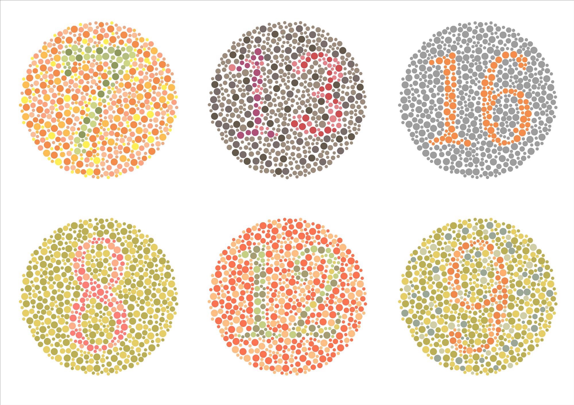
Is your blue the same as mine? Though we all use the same terms to describe a set of colors, the way each color is perceived will be unique to the individual.

Image Credit: Maria Vonotna/Shutterstock.com
Being trichromats, humans have cone photoreceptors in the retina that are sensitive to short, medium, and long wavelengths of light. Short/blue cones show maximum sensitivity at λB 430 nm, green/medium cones at λG 530 nm, and long/red cones at λR 560 nm. Stimulating these cones in complex patterns creates a visual spectrum between axes of red-green and blue-yellow.
Visual transduction process
Humans also possess rod photoreceptors which allow for non-color vision in dim light. Rods and cones are comprised of four functional regions: the specialized inner and outer segments, as well as a nuclear region and synaptic region. The outer segment of the rod, or cone, houses discs of photoreceptive membrane; connecting to the nuclear and synaptic regions via the inner segment.
Discs of photoreceptive membrane form as dense stacks, internalized and separated from the membrane in rods. In cones, the photoreceptive discs remain as foldings of the plasma membrane; thought to offer a larger surface area for processes such as chromophore transfer, as well as aiding rapid calcium dynamics. By stimulating these discs with a photon of light, visual information can be transmitted from synaptic terminals to higher centers in the brain.
Both rod and cone photoreceptors use phototransduction to convert stimuli into electrical impulses. Light stimulated activation of a G-protein-coupled receptor in the photoreceptive discs, activates cyclic GMP phosphodiesterase. By hydrolyzing cyclic GMP, the phosphodiesterase is able to reduce the concentration and cause cyclic-nucleotide-gated ion channels in the plasma membrane to close. This results in cell hyperpolarization and a low rate of transmitter release.
The visual information is then communicated to the thalamus by parvocellular layers, where the lateral geniculate nucleus processes and relays the signal to the primary visual cortex V1.
Genetic variation and visual perception
Any slight differences in the genes involved in the visual transduction response can mediate differences in color perception. Color blindness is a common condition and a prime example of how genetics can influence the perception of color. ~1 in 12 men and ~1 in 200 women are affected by color vision deficiencies globally. As well as genetic variation, an individual’s perception of color can be altered by acquired abnormalities in the visual system (including the retina, optic nerves, optic tract, and visual cortex) and disease.
Cones for medium and long wavelengths of light are carried on the X-chromosome; with short-wavelength cones coded on chromosome 7. The genes responsible for long and medium wavelength cones are believed to undergo frequent homologous recombination, contributing to the variability of color perception. Due to the sex-linked nature of these genes, karyotypic males are more susceptible to red-green color blindness.
On the X chromosome, the OPN1MW and OPN1LW genes encode opsin pigments in the medium and long-wavelength cones, respectively. In red-green color blindness, the most common form, affected individuals lack functional medium and/or long-wavelength cones. While genetic variation in these coding regions is most often due to errors in homologous recombination, single nucleotide polymorphisms can also be responsible.
If the individual only loses functional medium wavelength cone opsins, due to variation in the OPN1MW genes, the condition is termed deuteranopia. Protanopia is also red-green color blindness but is due to mutations in OPN1LW leading to non-functional long wavelength cones.

Image Credit: eveleen/Shutterstock.com
Loss of the color blue
Tritanopia is a relatively uncommon condition, where there are non-functional short-wavelength cone opsins, due to mutations in the OPN1SW gene. There are six known inherited mutations associated with this condition; one of which encodes a progressive degeneration of small wavelength cones. For these individuals, their perception of blue is lost with age.
Blue-yellow color vision deficiencies have also been associated with old age, with senescence of the lens thought to influence the ability to absorb short wavelengths. High UV-B exposure has also been linked to elevated ocular aging and lens brunescence. In regions of higher elevation, as well as locations closer to the equator, the average levels of UV-B exposure are relatively high. In these geographical locations, there is often a lack of verbal distinction for the color terms green and blue. It has been proposed that this lack of distinction between blue and green is due to premature senescence of the lenses.
Despite initial theories proposing that brunescent lenses impair perception of short wavelengths/blue colors. The physiological mechanism behind the regional lexicon of terms for blue and green has not yet been identified. Current opinion suggests there is a third mechanism linking accelerated lens brunescence and an absence of blue.
Sources:
- Fu Y. Phototransduction in Rods and Cones. (2010) Webvision: The Organization of the Retina and Visual System [Online]. Salt Lake City (UT): University of Utah Health Sciences Center. Available at: https://www.ncbi.nlm.nih.gov/books/NBK52768/ (Accessed on 1 November 2021).
- Klaus, C., et al. (2019) Multi-scale, numerical modeling of spatio-temporal signaling in cone phototransduction. PLoS One. doi:10.1371/journal.pone.0219848.
- Goulart, V.D.L.R., et al. (2017) Medium/Long wavelength sensitive opsin diversity in Pitheciidae. Sci Rep. doi:10.1038/s41598-017-08143-2.
- Pasmanter, N., & Munakomi, S., (2021) Physiology, Color Perception. [Online]. StatPearls Treasure Island (FL): StatPearls Publishing. Available at: https://www.ncbi.nlm.nih.gov/books/NBK544355/ (Accessed on 1 November 2021).
- Inherited Colour Vision Deficiency. [Online]. Colour blind awareness. Available at: www.colourblindawareness.org/.../ (Accessed on 1 November 2021).
- Colour Blindness. [Online]. Colour blind awareness. Available at: https://www.colourblindawareness.org/colour-blindness/ (Accessed on 1 November 2021).
- Genetics Home Reference. (2020) Colour vision deficiency [Online] MedlinePlus. Available at: https://medlineplus.gov/genetics/condition/color-vision-deficiency/ (Accessed on 1 November 2021).
- Werner, J.S. (2016) The Verriest Lecture: Short-wave-sensitive cone pathways across the life span. J Opt Soc Am A Opt Image Sci Vis. doi:10.1364/JOSAA.33.00A104.
- Hardy, J.L., et al. (2005) Color naming, lens aging, and grue: what the optics of the aging eye can teach us about color language. Psychol Sci. doi:10.1111/j.0956-7976.2005.01534.x.
- Neitz, M., et al. (2020) Tritan color vision deficiency may be associated with an OPN1SW splicing defect and haploinsufficiency. J Opt Soc Am A Opt Image Sci Vis. doi:10.1364/JOSAA.381919.
Further Reading