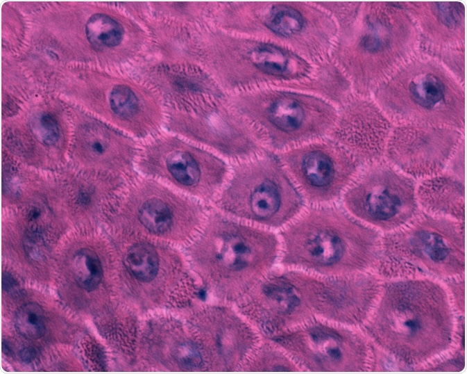
The skin provides an essential first line of defense protecting the body against potentially threatening substances in the environment.

Image Credit: Jose Luis Calvo/Shutterstock.com
To function properly the skin’s outer layer, the epidermis, must undergo the homeostatic processes of proliferation, differentiation, and apoptosis. The keratinocyte stem cells are vital in this regulatory cycle.
While their full function has yet to be determined, numerous studies have implicated their role in homeostatic skin processes.
Keratinocyte stem cells are found in the microenvironment of the basal epidermis, as well as in the adult hair follicle, and sebaceous glands. Because of their protection from apoptosis, a form of regulated cell death, they are prime candidates for the origin of skin cancer.
Below we discuss the role keratinocyte stem cells play in differentiation/proliferation and apoptosis, as well as how they are involved in the initiation and development of cancer.
Differentiation/proliferation in interfollicular stem cells
The interfollicular stem cell epidermis is under a continuous cycle of regeneration. This process is dependent on the proliferation of the interfollicular stem cells, a subset of the basal keratinocytes.
The epidermal proliferative unit (EPU) model dictates there is a proliferative heterogeneity in the basal layer of the epidermis, where the division of a single stem cell produces a stem cell daughter and a non-stem committed progenitor cell, known as a transit-amplifying (TA) cell.
While research has helping to uncover a number of markers that have assisted in isolating and characterizing enriched keratinocyte subpopulations, a robust method of epidermal stem cell identification and their niche remains elusive.
What we do know is that the subpopulation of keratinocyte stem cells, the IF cells, are responsible for generating new cells in the epidermis.
Differentiation in the hair follicle
Keratinocyte stem cells also exist in the hair follicle. Here they mediate the various stages of growth, apoptosis-mediated regression, and quiescence. Hair specific stem cells are located in the bulge region of the hair follicle epithelium.
The stem cells regenerate the new hair shaft from within this bulge region, as it provides a unique environment for restricted differentiation of the different types of adult stem cells. Both melanocyte stem cells and stem cells with neural crest characteristics are located in the bulge area.
Many studies have demonstrated the differentiation of stem cells in this location of the hair shaft, confirming that it is a critical zone for this homeostatic process.
Apoptosis
Because keratinocyte stem cells are located within a special microenvironment they retain their “stem” characteristics. Interfollicular keratinocyte stem cells that are located in the epidermis’s basal layer have access to the abundance of proteins and growth factors that are available in the extracellular matrix.
When exiting the niche, the basal keratinocytes relocate into the suprabasal layers where different microenvironmental factors impact their activity.
Usually, upon exiting the basal niche the basal keratinocytes undergo terminal differentiation which is regulated by a rising number of signals.
Keratinocytes become committed to differentiation as the notch receptors in suprabasal cells bind to the notch ligands expressed in basal keratinocytes.
This binding that initiates differentiation promotes the change between keratins 5/14 and K1/10, and although these markers are involved in differentiation regulation, they are also involved in triggering the process of programmed cell death, known as apoptosis.
Research has suggested that in the presence of different regulatory inputs, the basal cells are triggered to leave the niche and begin the process of cell death.
Skin cancer
Research is mounting to support the hypothesis that keratinocyte stem cells are the targets in skin carcinogenesis.
Characteristics of the stem cells, such as that they do not go through the genome protecting process of asymmetric chromosome segregation, and that they are incredibly resistant to apoptosis, means that they are susceptible to oncogenic mutations.
These mutations that occur in the absence of apoptosis and asymmetric chromosome segregation are associated with the initiation of skin cancer.
Recent studies have demonstrated that keratinocyte stem cells are characterized by their high resistance to apoptosis. In addition, research has uncovered that survivin that is found in high levels in squamous cell carcinoma is caused by chronic UV exposure and can be traced back to keratinocyte stem cell origination.
Finally, studies have also found that survivin and its antiapoptotic isoforms are expressed mostly in keratinocyte stem cells. This evidence highlights the link between keratinocyte stem cells and the initiation and development of skin cancer.
Summary
Keratinocyte stem cells exist in the microenvironment of the basal epidermis in three distinct locations, as interfollicular stem cells of the basal layer, as hair follicle stem cells of the bulge, and as the sebaceous gland stem cells that are found beneath the hair shaft orifice.
These stem cells are involved in the homeostatic processes of proliferation, differentiation, and apoptosis which maintain the skin’s normal functions. In addition, numerous studies have implicated keratinocyte stem cells in the establishment and development of skin cancer.
Sources:
- Alonso, L. and Fuchs, E. (2003). Stem cells of the skin epithelium. Proceedings of the National Academy of Sciences, 100(Supplement 1), pp.11830-11835. https://www.pnas.org/doi/10.1073/pnas.1734203100
- Pincelli, C. and Marconi, A. (2010). Keratinocyte stem cells: Friends and foes. Journal of Cellular Physiology, 225(2), pp.310-315. https://www.ncbi.nlm.nih.gov/pubmed/20568222
- Singh, A., Park, H., Kangsamaksin, T., Singh, A., Readio, N. and Morris, R. (2012). Keratinocyte Stem Cells and the Targets for Nonmelanoma Skin Cancer†. Photochemistry and Photobiology, 88(5), pp.1099-1110. https://www.ncbi.nlm.nih.gov/pmc/articles/PMC3357445/
- Wang, E. (2019). Immune cells and the epidermal stem cell niche. Advances in Stem Cells and their Niches, pp.193-218. https://www.sciencedirect.com/science/article/pii/S2468509719300065
Further Reading
Last Updated: Jul 18, 2022