The top-down approach is a strategy for processing information where a whole system is broken down into component parts, rather than being built up through the contrasting bottom-up approach.

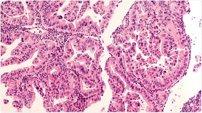
Top-down tissue microproteomics have been performed on benign, tumor, and necrotic-fibrotic areas of ovarian cancer biopsies. Credit: David Litman/ Shutterstock.com
This directional pathway for analysis and fabrication has been employed in a variety of fields from software development to economic theory. In the life sciences, the top-down approach has led to the development of new methodologies for protein analysis, providing greater detail of the proteoform within its natural context.
The ability to define protein profiles has important implications for future drug development and producing new biomarkers of disease. When we think of the manufacturing of biomedical devices it is natural to assume that they are all built through a bottom-up approach by piecing together components. However, modern devices, such as microfluidic biosensors, are being fabricated by a top-down approach that can easily define the small structures needed.
A top-down approach to proteomics
The top-down approach is being employed in the field of proteomics for the large-scale study of proteins. The conventional bottom-up approach to protein identification requires the digestion of proteins into component peptides, which are then introduced to a mass spectrometer. The fragmentation-based analysis detects the peptides which are used to infer the original protein.
The top-down approach introduces proteins directly into the mass spectrometer with a proteome map formed before analysis. The original proteins no longer have to be assembled through computational methods. Moreover, this type of analysis provides a full characterization of the proteoform without the loss of detail that can occur because of the digestion process.
The ability to provide more detailed proteomics data will be invaluable for the development of protein-based drugs. To utilize proteins for targeted therapy, a greater understanding of proteome characterizations such as post-translational modification must occur. The top-down methodology enables this by mapping large proteome complexes whilst preserving post-translationally modified protein forms.
Top-down approach for biomarker identification
The top-down approach to proteomics has also supplied the ability to characterize the specific protein profiles of different cancer regions. Top-down tissue microproteomics was performed on a benign, tumor, and necrotic-fibrotic areas of ovarian cancer biopsies.
The region-specific cellular localization of proteins and their post-translational modifications can be used to distinguish between tumor and benign zones. The method identified proteoform markers of ovarian cancer and the extracted proteins from each region produced several potential biomarkers of the disease.
The top-down approach can therefore be utilized in identifying diagnostic biomarkers that would be missed through conventional proteomics methodologies that rely on reference databases of proteins.
Top-down approach for fabricating microfluidic biosensors
Biosensors are used to detect biomolecules such as enzymes by forming a measurable signal, which indicates the identification and concentration of the target biomolecule. A common example of this technology is for glucose detection, with glucose biosensors providing an important tool for diabetes management.
Microfluidic biosensors can detect the target biomolecule in small sample volumes by the interaction between the analyte and the mechanism of signal transduction on a microarray. A top-down approach is being applied to the fabrication of microfluidic biosensors that is faster than the bottom-up approach which creates biosensors through the integration of material components one at a time.
Though bottom-up manufacturing of biosensors may be advanced through self-assembly, the most frequently used top-down method manufactures microfluidic biosensing devices by defining channels on a blank substrate through lithography. The multiple fluidic channels required for the detection of the target biomolecule throughout the device are therefore defined from the larger surface structure.
Sources:
- Catherman, A.D. et al. 2014. Top Down Proteomics: Facts and Perspectives, Biochemical and Biophysical Research Communications , 21, pp. 683-693. https://www.ncbi.nlm.nih.gov/pmc/articles/PMC4103433/
- Toby, T.K. et al. 2016. Progress in Top-Down Proteomics and the Analysis of Proteoforms, Annual Review of Analytical Chemistry, 9, pp. 499-519. https://www.ncbi.nlm.nih.gov/pmc/articles/PMC5373801/
- Suiti, N. & Kelleher, N.L. 2007. Decoding protein modifications using top-down mass spectrometry, Nature Methods, 4, pp. 817-821. https://www.ncbi.nlm.nih.gov/pmc/articles/PMC2365886/
- Delcourt, V. et al. 2017. Combined Mass Spectrometry Imaging and Top-down Microproteomics Reveals Evidence of a Hidden Proteome in Ovarian Cancer, EBioMedicine, 21, pp. 55-64. https://www.sciencedirect.com/science/article/pii/S2352396417302293
- Prakash, S. et al. 2012. Theory, fabrication and applications of microfluidic and nanofluidic biosensors, Philosophical Transactions of the Royal Society A, 370, pp. 2269-2303. http://rsta.royalsocietypublishing.org/content/370/1967/2269
Further Reading
Last Updated: Feb 2, 2021