As the name suggests, mass spectrometry (MS) is an analytical technique that measures the mass of a sample. More precisely, MS measures the mass-to-charge ratio of the ions generated by the ionization of the molecules present in a given sample.

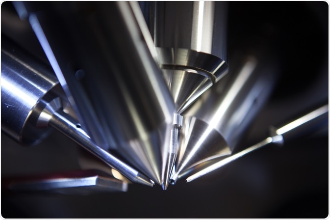
Mass Spectrometry. Image Credit: Intothelight Photography/Shutterstock.com
In reality, mass spectrometry does much more than just measuring the mass. In addition to providing information about the exact mass of a sample and its fragments, the technique can also analyze the elemental composition and isotopic distribution.
Almost every substance can be analyzed by mass spectrometry, from low-molecular-weight molecules (i.e., drugs, additives, contaminants) to macro-and biomolecules, such as polymers, proteins, and DNA. A powerful aspect of mass spectrometry is that not only it can be used independently, but it can also be coupled to other techniques such as gas or liquid chromatography (GC-MS and LC-MS respectively).
The information obtained can be used for the structural elucidation of chemical compounds, finding application in several fields, ranging from pharmaceutical research, food industry, and environmental science, just to name a few. For instance, mass spectrometry has a pivotal role in forensic science, with many drugs being identified with the analysis of urine, hair, and blood.
General principles and instrumentation
In an MS experiment the sample – either solid, in solution, or the gas phase – is ionized, causing the molecules to become charged and in some cases break up in fragments. These charged ions are accelerated in an electric field and then deflected by a magnetic field.
This last step enables the separation of ions according to their mass-to-charge ratio (m/z). Ions with different m/z ratios will have different deflections. Eventually, the ions reach a detector where an electric signal is converted into what we know as the mass spectrum.
Therefore, all mass spectrometers can be broken down into four main components. The first one is the ion source. Depending on the sample type there are different ionization modes, with the most common being electrospray ionization (ESI) and matrix-assisted laser desorption/ionization (MALDI).
The second component is the mass analyzer that separates the ions according to their m/z ratio. Among the most common alternatives, we find quadrupole mass analyzers and ion traps. Time of flight (TOF) is another popular type of mass analyzer. With TOF, ions formed in the source are accelerated by an electric field with known potential, giving the ions the same kinetic energy. As a consequence, the velocity of the ions depends on their m/z ratio, and so does the time required to reach the detector. Therefore, time can be used as a parameter to measure the ions’ mass.
The third component is the detector to measure the abundances of the separated ions as an electrical signal. They are typically either electron multipliers or array detectors, with the latter being used to detect ions simultaneously and to increase sensitivity.
The last component is a recording device that converts the detector’s signal into processable data, allowing for the generation of mass spectra, which are the plot of the ion signals as a function of the mass-to-charge ratio.
Beyond conventional analysis - Mass Spectrometry Imaging
Mass spectrometry is also used for experiments that go beyond the conventional analysis of known or unknown samples. It is possible to investigate the spatial distribution of molecular species thanks to mass spectrometry imaging (MSI).
This technique, which is becoming more established in clinical practice and the pharmaceutical industry, is a valuable analysis tool for the characterization of a biological specimen, such as cells or tissues. MSI can image thousands of molecules (e.g., metabolites, proteins, glycans, etc.) in a single experiment without labeling.
Following careful sample preparation, the user chooses an area defining an (x, y) grid over the surface of the sample. The molecules on the surface of the sample are then ionized and a mass spectrum is collected at each pixel on the section.
After collecting the spectra, software can be used to select an individual m/z value, and the intensity is extracted from each pixel. These intensities are then combined into a heat map image representing the relative distribution of the species on the sample’s surface.
The identity of a specific m/z value can be determined by tandem MS (MS/MS) fragmentation on ions from each pixel, using the fragments to reconstruct the structure of the unknown molecule. Alternatively, molecules can also be identified via their intact mass by accurate mass-matching to databases of known molecules.
TOF-SIMS for surface analysis
In the last two decades, time-of-flight secondary ion mass spectrometry (TOF-SIMS) has emerged as one of the most important and versatile techniques for surface analysis in advanced materials research. TOF-SIMS can provide information on the chemical composition of a surface.
The surface is bombarded with a pulsed ion beam (primary ions). The resulting atomic collisions transfer the primary ion energy to the target atoms, initiating a collision cascade which ultimately generates secondary species. A small percentage of these secondary species comes off as either positive or negative ions, which are extracted into an analyzer where their mass is determined.
Since a TOF-SIMS spectrum consists of numerous peaks arising from the fragmentation of the molecules on the surface, the technique provides a useful fingerprint of the surface chemical composition.
Conclusions
Mass spectrometry plays a central role in the analytical field and there are no doubts about how important and versatile it can be. There are continuous technological developments for more accurate analyzers, sensitive detectors, and efficient programs for data processing. Moreover, with imaging techniques becoming more and more established, future applications of mass spectrometry are limitless.
References
- López-Ruiz, R., Romero-González, R. & Garrido Frenich, A. (2019). Ultrahigh-pressure liquid chromatography-mass spectrometry: An overview of the last decade. TrAC Trends in Analytical Chemistry, 118, 170-181.10.1016/j.trac.2019.05.044
- Johnstone, R. a. W. & Rose, M. E. 1996. Mass Spectrometry for Chemists and Biochemists, Cambridge, Cambridge University Press.10.1017/CBO9781139166522
- Buchberger, A. R., Delaney, K., Johnson, J. & Li, L. (2018). Mass Spectrometry Imaging: A Review of Emerging Advancements and Future Insights. Anal Chem, 90, 240-265.10.1021/acs.analchem.7b04733
- Sodhi, R. N. (2004). Time-of-flight secondary ion mass spectrometry (TOF-SIMS):--versatility in chemical and imaging surface analysis. Analyst, 129, 483-7.10.1039/b402607c
Further Reading
Last Updated: Sep 13, 2022