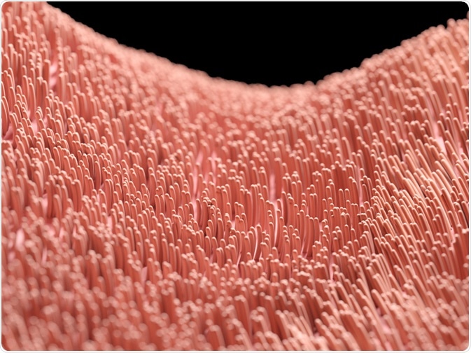
Cilia are microtubule-based cellular protrusions that serve a wide range of functions, and defects in its development or structure have pathological consequences.
Unicellular organisms rely on cilia for locomotion, feeding, and sensation, and many eukaryotic cells possess none or a few cilia; however, some cells possess multiple cilia that beat in a polarised and synchronized fashion to carry out its functions.

Image Credit: SciePro/Shutterstock.com
Cilia structure and mechanism of beating
Cilia are anchored on the cell surface by a modified centriole structure called the basal body. The protrusion of the cilia extending from the basal body to the extracellular space is known as the axoneme, which is made up of nine pairs of microtubules organized circumferentially and enclosed by a specialized plasma membrane.
In the center of the microtubule ring lies a pair of central microtubules. Cilia motility is accomplished by the ring of microtubules and the central pair sliding against each other.
The sliding movement is constrained by proteins anchored at the basal bodies and between adjacent microtubule doublets, resulting in the bending of the cilium.
Types and functions of multiciliated cells (MCC)
In vertebrates, most cells develop a single, non-motile cilium for the regulation of signal transduction during development and homeostasis. Some specialized cells develop multiple cilia on their cell surface, which beat in a synchronized way primarily to drive the transport of liquid.
MCC is found in the spinal cord and ventricles of the adult brain. The coordinated beating of the cilia drives a bidirectional flow of cerebrospinal fluid (CSF) through the spinal cord and exchanging molecules between the brain and the spinal cord (Figure 2).
This is also important for neuronal migration. The function of the central nervous system depends on the controlled migration of young neurons and studies have found that the movement of young neurons is in parallel with the flow of CSF.
MCC plays an important role in governing the circulation of CSF and neurons in the central nervous system. Similarly, MCC is also found within the brain ventricular system, which is the CSF-filled cavities in the brain. CSF is circulated throughout the brain by the beating of MCC to nourish the brain.
In the airway, ventilation proposes a threat of infection to the human body. Throughout evolution, a variety of defense mechanisms have been developed, one of which involves the clearance of mucus by MCCs in the airway.
Mucus is an aqueous secretion that is produced by specialized cells in the respiratory system called goblet cells. Mucus moistens and protects the respiratory epithelium, cells that line the airway.
Since pathogens and foreign particles such as dust trapped on mucus needs to be cleared, therefore, the respiratory epithelial cells, which possess around 200 cilia each, use cilia beating to target movement of mucus upwards of the respiratory tract to propel pathogens out of the system.
Moreover, multiciliated cells are also present in fallopian tubes, which are ducts in the female reproductive system that transport the egg from the ovary to the uterus. Propulsion of gametes and embryos depends on the interaction between muscle contractions, the beating of the cilia of MCCs, and the flow of tubal secretions.
Although the mechanism of transport and the relative importance of the three factors mentioned above are unclear, it is believed that cilia play a dominant role in propelling the transport of ovum from the ovary to the uterus, and sperms have been shown to bind to the ciliated areas of the outer lining of the fallopian tube.
Defects in MCCs
As MCCs play important functions in the nervous, respiratory and reproductive system, disruptions to the development of MCCs and thus the flow of fluids they facilitate lead to conditions such as chronic lung infections, increased risk of accumulation of CSF in the brain and decreased female fertility.
Primary cilia dyskinesia is an autosomal recessive genetic disorder that causes defects in cilia movements. It could affect the cells lining the respiratory tract, fallopian tube, and flagellum of sperm cells.
This is because the structure of the cilium is incomplete, and components of the cilium are missing during development. This results in unsynchronised or insufficient cilia beating.
One of the most common types of phenotype is the lack of central pair of microtubules in the cilium, making the cilium rotating instead of beating. There is currently no standardized treatment for this genetic disease. Instead, patients are given treatments to complications that arise from primary cilia dyskinesia.
Reference:
- Boutin, C. & Kodjabachian, L. (2019) Biology of multiciliated cells. The molecular and genetic basis of disease. Available from: http://www.sciencedirect.com/science/article/pii/S0959437X18300960.
- Brooks, E. R., & Wallingford, J. B. (2014). Multiciliated cells. Current biology: CB, 24(19), R973–R982. https://doi.org/10.1016/j.cub.2014.08.047
- Lyons, R., Saridogan, E., O. Djahanbakhch, O. (2006) The reproductive significance of human Fallopian tube cilia. Human Reproduction Update, 12(4), 363–372, https://doi.org/10.1093/humupd/dml012
- Ringers, C., Olstad, E. W. & Jurisch-Yaksi, N. (2020) The role of motile cilia in the development and physiology of the nervous system. Philosophical Transactions of the Royal Society B: Biological Sciences. 375 (1792), 20190156.
- Roy, S. (2017) Primary Ciliary Dyskinesia. Available from: http://www.rarediseasesindia.org/pcdyskinesia?tmpl=%2Fsystem%2Fapp%2Ftemplates%2Fprint%2F&showPrintDialog=1.
Further Reading
Last Updated: Jan 28, 2021