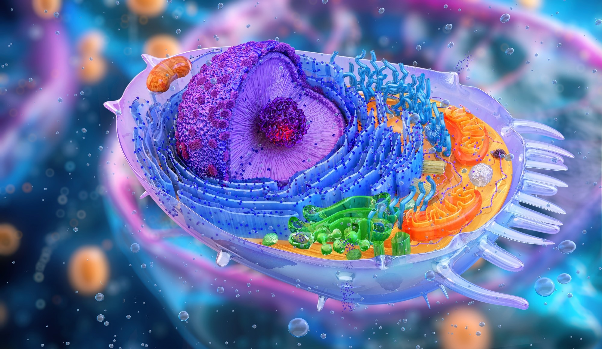
Protein synthesis refers to the process by which cells construct proteins. It's a fundamental cellular mechanism for all living organisms, as proteins carry out enzymatic activities, structural support, transport, signaling, and immune defense.
 Image Credit: Corona Borealis Studio/Shutterstock.com
Introduction
The genetic information encoded in DNA provides information for protein synthesis. This information is first transcribed into messenger RNA (mRNA), which then serves as a template for translation, the process where ribosomes read the mRNA sequence and assemble amino acids into a polypeptide chain.
Comparing protein synthesis in prokaryotic vs. eukaryotic cells is essential for drug development. Many antibiotics target specific differences in prokaryotic proteins, allowing them to kill bacteria selectively without harming human cells.
The Steps of Protein Synthesis
Key Differences in Protein Synthesis
Overview
In prokaryotes like bacteria, transcription and translation can occur simultaneously in the cytoplasm, as there is no nuclear membrane to separate these processes.
In eukaryotic cells like human cells, transcription occurs in the nucleus, and the resulting mRNA is then transported to the cytoplasm for translation.
Additionally, eukaryotic mRNA often undergoes post-transcriptional modifications, such as splicing, capping, and polyadenylation, before it is exported from the nucleus. These modifications can regulate gene expression and increase mRNA stability.
Ribosomal Structure
Ribosomes, the cellular machines responsible for protein synthesis, are essential for all living organisms. Prokaryotic cells possess 70S ribosomes (where S refers to the Svedberg unit, which is a measure of the sedimentation coefficient), and they are smaller and simpler compared to eukaryotic ribosomes. These 70S ribosomes consist of two subunits: a 30S small subunit and a 50S large subunit. The 30S subunit contains a 16S ribosomal RNA (rRNA) molecule, while the 50S subunit harbors 23S and 5S rRNA molecules.
In contrast, eukaryotic cells have 80S ribosomes, which are larger and more complex and also consist of two subunits: a 40S (small) subunit and a 60S (large) subunit. The 40S subunit contains an 18S rRNA molecule, and the 60S subunit harbors 28S, 5.8S, and 5S rRNA molecules.
The unique structural features of prokaryotic ribosomes make them an ideal target for many antibiotics. These drugs can selectively bind to and inhibit the function of bacterial ribosomes without affecting eukaryotic ribosomes, leading to the death of the bacterial cell.1
mRNA Processing
Prokaryotic genes are generally organized in a continuous, uninterrupted manner, lacking introns, which are non-coding DNA sequences that interrupt the coding regions (exons) of eukaryotic genes. Since prokaryotic genes are already compact and efficient, there's no need for introns to be removed. This simplified structure allows for rapid transcription and translation, enabling prokaryotes to respond quickly to environmental changes.
In contrast, in eukaryotic genes, the primary RNA transcript, known as pre-mRNA, undergoes a process called splicing. During splicing, introns are excised, and exons are joined together to form a mature mRNA molecule. The splicing process is essential for generating functional mRNA and producing diverse protein isoforms from a single gene.
To further protect and stabilize their mRNA molecules, eukaryotic cells add a 5' cap and a poly-A tail to the mRNA transcript. The 5' cap, a modified guanosine nucleotide, is added to the 5' end of the mRNA molecule. It protects the mRNA from degradation by cellular enzymes and aids in the initiation of translation. The poly-A tail, a chain of adenine nucleotides, is added to the 3' end of the mRNA. It enhances mRNA stability, facilitates mRNA export from the nucleus, and plays a key role in translation termination.
Initiation Mechanisms
In prokaryotes, translation initiation depends on the Shine-Dalgarno sequence, which is typically located about eight nucleotides upstream of the start codon and is complementary to a region in the 16S rRNA of the small ribosomal subunit. The base pairing between the Shine-Dalgarno sequence and the 16S rRNA helps position the ribosome correctly on the mRNA, ensuring accurate translation initiation.
Eukaryotes, on the other hand, utilize the Kozak sequence to start translation. The Kozak sequence surrounds the start codon (AUG) and has a consensus sequence of (GCC)GCCRCCAUGG, where R represents a purine (i.e., adenine and guanine). The Kozak sequence enhances the efficiency of translation initiation by providing a favorable context for the ribosome to recognize the start codon.
Eukaryotic and Prokaryotic Differences in Transcription and Translation
Applications and Implications
The differences in ribosomal structure reflect the evolutionary divergence between prokaryotes and eukaryotes. Over billions of years, these two major groups of organisms have evolved distinct cellular machinery, including ribosomes.
These differences in ribosomal structure have allowed for the development of antibiotics that can selectively target bacteria without harming the host organism.
Many antibiotics, such as aminoglycosides and macrolides, bind to specific sites on the prokaryotic ribosome, interfering with its function.2,3
These antibiotics can inhibit protein synthesis by disrupting the formation of peptide bonds, preventing the binding of tRNA molecules, or causing premature termination of translation. Since eukaryotic ribosomes have a different structure, these antibiotics do not significantly affect them, making them effective against bacterial infections.
Advantages and Limitations of Each System
Prokaryotic protein synthesis offers the advantage of rapid production due to the coupled nature of transcription and translation. This simplicity makes them ideal for laboratory applications and large-scale protein production.
Escherichia coli expression systems are particularly ideal for producing therapeutic proteins on a lab scale and in industry due to its low cost and simplicity of cultivation.4 Similarly, Bacillus subtilis strains engineered for improved protein production exhibit advantages in time and cost compared to competing systems.5
While slower, eukaryotic protein synthesis provides a higher degree of regulation than prokaryotic systems, allowing for precise control of gene expression and protein production.
Eukaryotic proteins have also undergone a wide range of taxon-specific post-translational modifications, such as glycosylation and phosphorylation, which are essential for proper function.
Do Your Genetics Determine Your Intelligence?
Conclusion
Understanding the differences in protein synthesis among prokaryotic and eukaryotic organisms has profound implications for scientific research, medical applications, and biotechnology.
By studying these fundamental processes, scientists can gain insights into the evolution of life, develop novel therapeutic strategies, and advance our understanding of cellular biology.
References
- Wilson, D. (2013). Ribosome-targeting antibiotics and mechanisms of bacterial resistance. Nature Reviews Microbiology, 12, 35-48. https://doi.org/10.1038/nrmicro3155.
- Kaul, M., Barbieri, C., & Pilch, D. (2004). Fluorescence-based approach for detecting and characterizing antibiotic-induced conformational changes in ribosomal RNA: comparing aminoglycoside binding to prokaryotic and eukaryotic ribosomal RNA sequences. Journal of the American Chemical Society, 126 11, 3447-53. https://doi.org/10.1021/JA030568I.
- Sothiselvam, S., Neuner, S., Rigger, L., Klepacki, D., Micura, R., Vázquez-Laslop, N., & Mankin, A. (2016). Binding of Macrolide Antibiotics Leads to Ribosomal Selection against Specific Substrates Based on Their Charge and Size. Cell reports, 16 7, 1789-99. https://doi.org/10.1016/j.celrep.2016.07.018.
- Kamionka, M. (2011). Engineering of Therapeutic Proteins Production in Escherichia coli. Current Pharmaceutical Biotechnology, 12, 268 - 274. https://doi.org/10.2174/138920111794295693.
- Pohl, S., Bhavsar, G., Hulme, J., Bloor, A., Misirli, G., Leckenby, M., Radford, D., Smith, W., Wipat, A., Williamson, E., Harwood, C., & Cranenburgh, R. (2013). Proteomic analysis of Bacillus subtilis strains engineered for improved production of heterologous proteins. PROTEOMICS, 13. https://doi.org/10.1002/pmic.201300183.
Further Reading
Last Updated: Mar 5, 2025