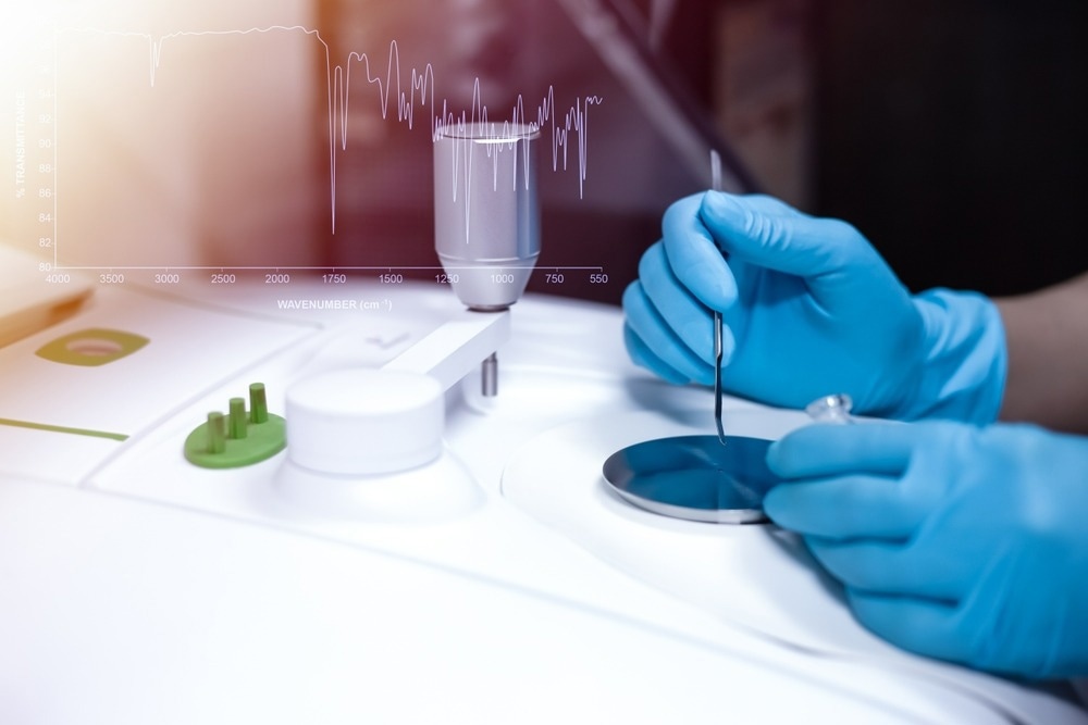
Trace elements are broadly defined as those with a concentration of less than 100 ppm or 100 mg per kg, with ultratrace elements with a concentration below 1 ppm, though these thresholds may vary by field and industry.

Image Credit: S. Singha/Shutterstock.com
Clinical diagnosis, environmental monitoring, and numerous other applications depend on accurate trace element analysis, for which spectroscopy-based techniques are popular owing to their wide adaptability, high throughput rate, and reliability. This article will discuss some of the methods of trace element spectroscopy and their applications in the sciences.
Introduction to Trace Elements and Their Importance
Elemental analysis is concerned with the quantification of elements within a sample, not the determination of molecular structure or any other higher-order arrangement of atoms. Ubiquitous elements, such as carbon within organic samples or silicon in rocks, are therefore not typically usefully assessed by spectroscopic methods of elemental analysis unless one wishes to know these quantities for specific research purposes. Instead, spectroscopy techniques are used for the elemental analysis of trace elements present in a wide range of samples, such as heavy metals in water, soil, rocks, or living tissue.
Fundamentals of Spectroscopy for Elemental Analysis
Spectroscopy is the study of the absorption and/or emission of light from matter, for which there are several mechanisms. The most simple and probably most widely employed type of spectroscopic elemental analysis technique is atomic adsorption spectroscopy (AAS), wherein the sample is firstly atomized and then exposed to incident light.
Particular elements are known to absorb light of specific wavelengths, and therefore a detector placed on the opposite site of the sample allows researchers to determine quantitatively which elements are present by the lessened intensity of absorption at these wavelengths. Similarly, atomic emission spectroscopy (AES) first atomizes the sample and then exposes it to incident radiation, in this case detecting photon emissions caused by excitation.
Inductively Coupled Plasma Optical Emission Spectrometry (ICP-OES)
Inductively coupled plasma-optical emission spectroscopy (ICP-OES) (or atomic emission spectroscopy (ICP-AES)) utilizes the principles of AES as described above and employs an inductively coupled plasma torch to atomize the sample. The torch is typically argon gas heated by inductive coupling using electrical coils, reaching temperatures approaching or exceeding 10,000 K that are capable of atomizing a wide range of sample types, which are usually delivered into the flame as a mist.
Since the spectral lines generated by emissions from specific elements are extremely narrow, numerous elements without overlapping signals can be simultaneously quantified. This is particularly useful where a broad range of trace elements are expected, such as environmental soil samples or engine oil samples during mechanical failure diagnosis. Prior to analysis by ICP-OES, a calibration run is typically performed using control samples containing increasing quantities of the elements of interest, obtaining a concentration to spectral intensity dose-response curve against which the intensity observed of the sample can be compared.
Inductively Coupled Plasma Mass Spectrometry (ICP-MS)
Inductively coupled plasma-mass spectrometry (ICP-MS) utilizes the same type of inductively coupled plasma torch as ICP-OES to atomize the sample, but in this case is in tandem with mass spectrometry apparatus. During atomization, the sample cloud is also ionized and, therefore, bears a charge. Ions are directed by magnetic fields within the mass spectrometry apparatus at a rate dependent on the ions' mass-to-charge ratio, which can then be used to infer their character.
Compared to ICP-OES, ICP-MS is a more sensitive order of magnitude, comfortably detecting trace elements well into the parts per trillion range. This provides numerous applications for which ICP-OES lacks sufficient sensitivity, such as in forensics, toxicology, and the pharmaceutical sciences.
X-Ray Fluorescence (XRF) Spectroscopy
X-ray fluorescence (XRF) spectroscopy operates similarly to AES spectroscopy in that it relies on the detection of emitted photons from a material that has been excited by incident light. In this case, however, X-rays or, in some cases, gamma rays are utilized, which are capable of ejecting electrons from the inner orbitals of the atom and causing electrons in higher orbitals to "fall" into the lower orbital to fill the hole.
The energy difference between these orbitals is emitted as a photon of characteristic wavelength to the atom from which it is emitted. XRF spectroscopy devices are available in a wide range of profiles for a range of specific applications, from small handheld devices suitable for fieldwork to huge apparatus into which large engineered materials can be placed.
One major advantage of the technique is that it is non-destructive to non-living materials, and thus glass, metals, ceramics, and other building materials are regularly checked for contaminants during manufacturing by XRF spectroscopy, and the technique is used non-destructively in forensic and art science investigation.
Laser-Induced Breakdown Spectroscopy (LIBS)
An emerging method of micro-destructive elemental analysis is known as laser-induced breakdown spectroscopy (LIBS), which utilizes short laser pulses to atomize and excite the sample, whereupon AES techniques are used to collect the emitted light and produce a spectrograph.
The technique was developed in the 2000s by the US Army Research Laboratory, which was concerned primarily with developing a method of analyzing hazardous samples such as explosive residues. The technique has since become useful in the analysis of samples in the field. It is used mainly for the high-speed identification of metals and plastics, such as in manufacturing, environmental analysis, the sorting of recycled materials, and so on.
Sources
- Bulska, E., & Ruszczyńska, A.. (2017). Analytical Techniques for Trace Element Determination. Physical Sciences Reviews, 2(5). https://doi.org/10.1515/psr-2017-8002
- Brouwer, P. (2010). Theory of XRF. https://home.iiserb.ac.in/~ramyasr/files/Manuals/XRF.pdf
- Anabitarte, F., Cobo, A., & Lopez-Higuera, J. M.. (2012). Laser-Induced Breakdown Spectroscopy: Fundamentals, Applications, and Challenges. ISRN Spectroscopy, 2012, 1–12. https://doi.org/10.5402/2012/285240
Further Reading
Last Updated: Dec 13, 2023