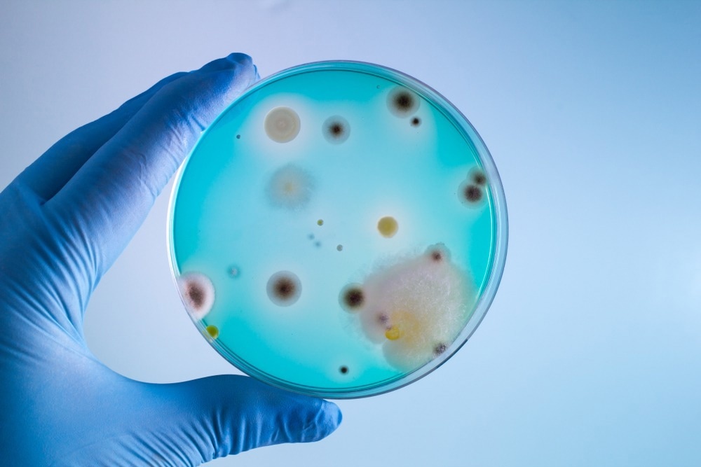
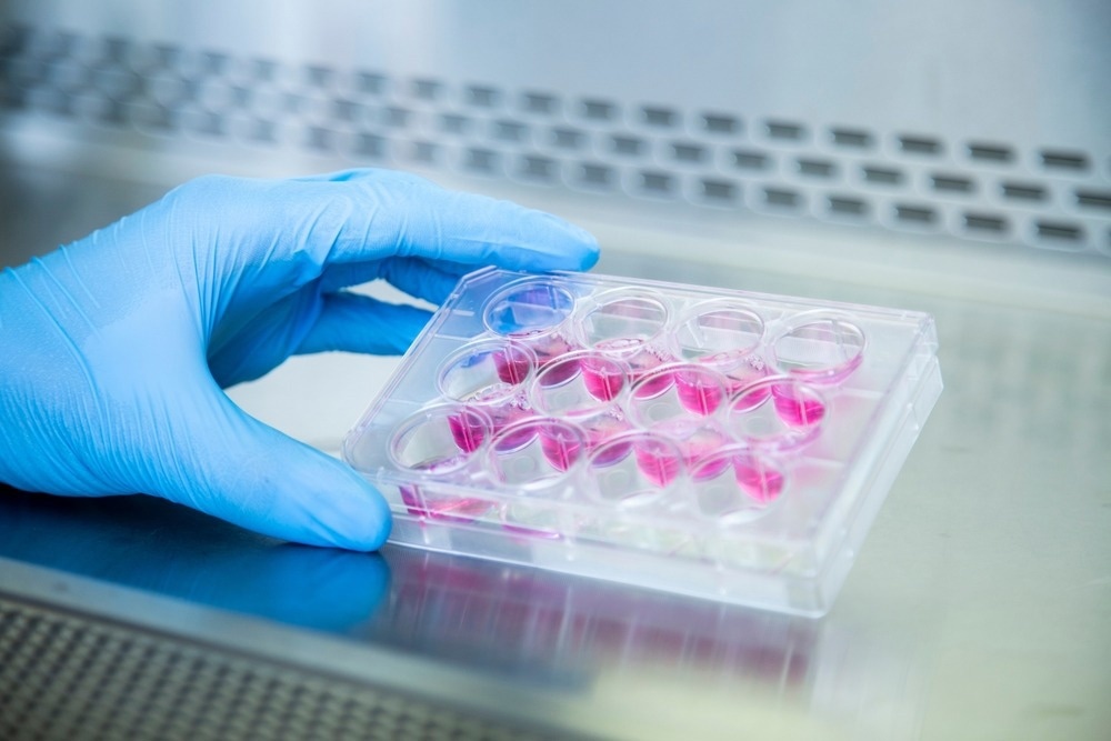
Cell culture contamination is a common problem encountered by cell biologists and laboratory technicians and could have serious consequences on results.

Image Credit: angellodeco/Shutterstock.com
Cell culture can be contaminated by biological agents, such as bacteria, fungi, mycoplasma, and viruses, and chemical contaminants that include media, sera, endotoxins, and water. Since contaminations cause experimental delays and financial loss, effective measures must be undertaken to detect and prevent cell culture contamination.
Importance and Types of Cell Cultures in Biological Research
Cell culture experiments are commonly used in biotechnological production, biomedical research, pharmaceutical industries, regenerative medicine, and biomedical research. Owing to restrictive laws and implementation of the 3Rs, namely, replacement, reduction, and refinement for animal welfare, the use of animals for experimental purposes has decreased significantly along with an increase in cell line use.
There are different types of cell lines, including finite cell lines, continuous cell lines, immortalized cell lines, and stem cell lines. Finite cell lines have a slow growth rate and are obtained from primary cultures. Typically, cells belonging to this cell line are arrested in the G0, G1, or G2 phase after monolayer formation.
An immortalized cell line is derived from transformed or cancerous cells that have a rapid growth rate. This cell line quickly reaches higher cell density in culture and might form multilayers. Stem cells are partially or undifferentiated pluripotent cell types derived from multicellular organisms. These cells serve as multipotent precursors of many different cell types.
Each cell culture exhibits distinct properties associated with viability, morphology, genetic stability, and doubling time. Their maintenance and handling may require different culture conditions, media, and processing agents (e.g., antibiotics and detachment solutions).
It is extremely important to perform cell culture experiments with accuracy as they are prone to errors. Cell culture assays often encounter biological, chemical, and inter- and intra-specific cross-contamination and cell misidentification. These contaminants lead to fast changes in the pH of the growth medium and increased cell death or floating cells. Therefore, cell culture experiments must follow good cell culture practice (GCCP) as it would ensure reproducibility in in vitro experiments.
Different Types of Cell Culture Contaminants
Cell cultures are contaminated with different types of biological and chemical contaminants. Some of the key contaminants are discussed below:
Biological Contamination
Both bacteria and fungi are present ubiquitously in the environment. The microorganisms can rapidly colonize in a rich culture milieu. In the absence of antibiotics, bacterial colonies can be detected within a few days of contamination via visual inspection or microscopy.
Mycoplasmas are self-replicating and small, i.e., sized between 0.1 and 0.2 µm, organisms that belong to the class Mollicutes. It is difficult to detect mycoplasma contamination under a standard bright-field microscope owing to their small size. As a result, these contaminants often go undetected. Notably, mycoplasma lacks cell walls and is resistant to common antibiotics, such as penicillin and streptomycin, that are used in cell culture.
Virus contaminants present a more serious threat compared to other biological contaminants. This is due to the difficulty in detecting them and the unavailability of proper methods to revive the affected cell cultures. Certain virus contaminants can also integrate with the host cell genome and can lead to horizontal virus transmission across other cell lines.
Chemical Contamination
Non-living components that induce undesirable effects in cell culture are referred to as chemical contaminants. Some of the common chemical contaminants are water, media, sera, unsterile glass, and plasticware (e.g., storage bottles, culture tubes, and cell culture disks). Furthermore, impurities present in the carbon dioxide flow and residues from disinfectants used to sterilize incubators or hoods can act as potential chemical contaminants.
Endotoxins, derived from the outer cell membrane of many Gram-negative bacteria, are regarded as cell culture contaminants. High concentrations of heavy metals can be toxic to many cell types. Hence, solvents used to prepare culture media must be tested for the presence of heavy metals before use.
Inter- and Intra-Species Cross-Contamination
Cell lines can be contaminated with other unrelated cells from the same species (intra-species contamination) or other species (inter-species contamination). A rapidly dividing cell line could easily outgrow and replace the original culture.

Image Credit: OMER FARUK ERCEYLAN/Shutterstock.com
Methods to Detect Cell Culture Contaminants
Cellular contaminants can be detected via many approaches. Some of the common cell culture contamination detection methods are discussed below:
Visual inspection
Bacterial contamination can be visually detected by the change in turbidity. The presence of yeast can be suspected if the culture medium becomes cloudy or turbid. In contrast, molds develop branched mycelium that appear as furry clumps floating in the medium.
Microscopic observation
Bacterial colonies appear within a few days of contamination and these can be observed under microscopes. Electron microscopy is a universal, unbiased, and effective method to detect virus contaminants. Particularly, transmission electron microscopy (TEM) is used to visualize virus contaminants in cell culture samples.
Molecular Tools and Techniques
Some common tools and techniques used to detect biological contaminants are DNA staining by fluorochromes, such as Hoechst 33342 and 4′,6-diamidino-2-phenylindole (DAPI). Furthermore, advanced Fourier transform infrared (FITR) micro-spectroscopy methods, sensitive PCR-based assays, flow-cytometric methods, and colorimetric or strip-based mycoplasma detection assays are also used. Short tandem repeat (STR) profiling is used to detect cross-contamination and cell misidentification.
Recommendations to Avoid Contamination
Regular testing for cell culture contaminants is highly recommended as it will minimize falsified research results, the waste of research resources, and misleading publications. Many companies, including Thermo Fisher Scientific, Sigma-Aldrich, Lonza Biosciences, and SwiftDx, have developed detection kits that are highly sensitive and specific. These kits can be effectively used for early detection of cell culture contaminants.
Many commercial kits are based on the conventional polymerase chain reaction (PCR) method. Some of the commercial kits are equipped to detect more than 90 mycoplasma species. These kits use primers designed to target and amplify the highly conserved 16S rRNA coding region of the mycoplasma genome. Effective use of bactericidal components can eliminate mycoplasma infections.
Some of the standard practices that can reduce cell line contamination include the use of routine monitoring of cell culture, following standard aseptic techniques, maintaining a clean laboratory, strategic use of frozen cell repositories, and limited use of antibiotics. If required, handlers must use gloves, masks, and gowns to prevent potential contamination.
Sources
Weiskirchen, S. et al. (2023) A Beginner’s Guide to Cell Culture: Practical Advice for Preventing Needless Problems. Cells. 12(5). https://doi.org/10.3390/cells12050682
On the frontline of the fight against cell culture contamination. (2023). Nature. [Online] Available at: https://www.nature.com/articles/d42473-019-00306-1
Ryan, J. (2023) Understanding and Managing Cell Culture Contamination. [Online]
Ramos-Junior, P.L.J. et al. (2021) Detection of Microbiological Contamination in Cell Cultures and their Raw Materials: a 6 years report of this activity. Journal of Physics: Conference Series. 2606. doi:10.1088/1742-6596/2606/1/012017
Chen, X. et al. (2020) Authentication, characterization and contamination detection of cell lines, xenografts and organoids by barcode deep NGS sequencing. NAR Genomics and Bioinformatics. 2(3). https://doi.org/10.1093/nargab/lqaa060
Niehues, H. et al. (2020) Know your enemy: Unexpected, pervasive and persistent viral and bacterial contamination of primary cell cultures. Experimental Dermatology. 29(7), pp. 672-676. https://doi.org/10.1111/exd.14126
Pinheiro de Oliveira, T. F. et al. (2013) Detection of contaminants in cell cultures, sera and trypsin. Biologicals. 41(6), pp. 407-414. https://doi.org/10.1016/j.biologicals.2013.08.005
Mirjalili, A. et al. (2005) Microbial contamination of cell cultures: A 2 years study. Biologicals. 33(2), pp. 81-85. https://doi.org/10.1016/j.biologicals.2005.01.004
Further Reading