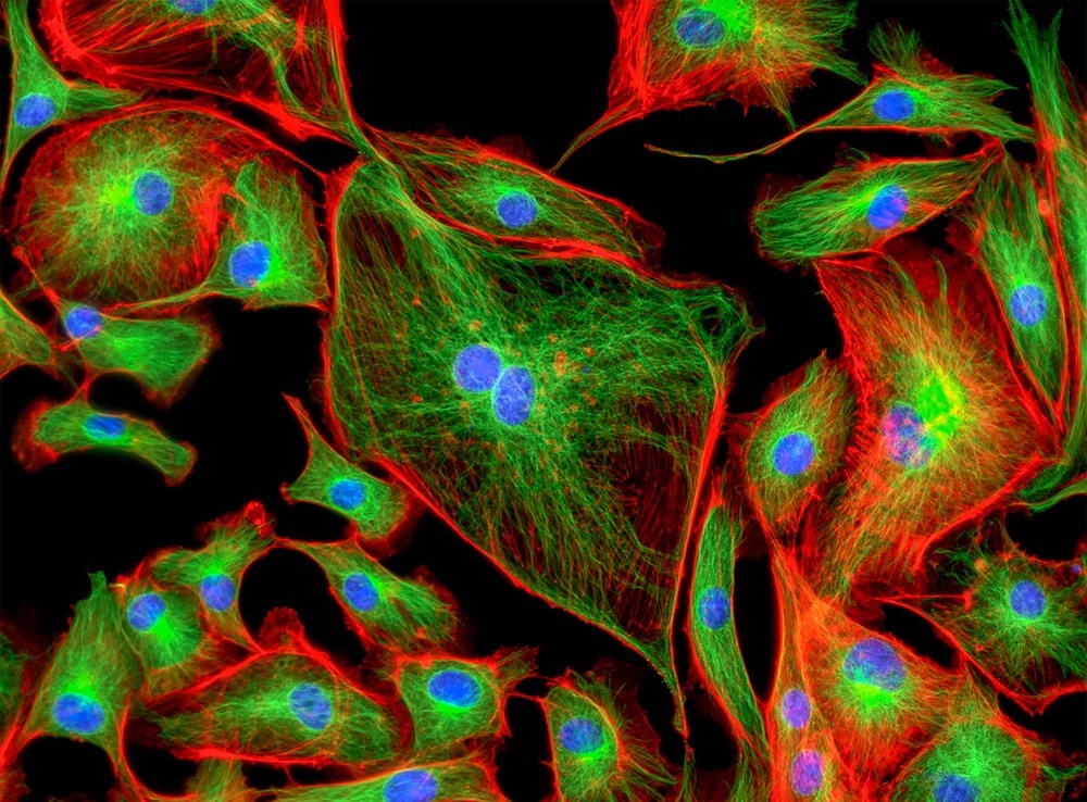
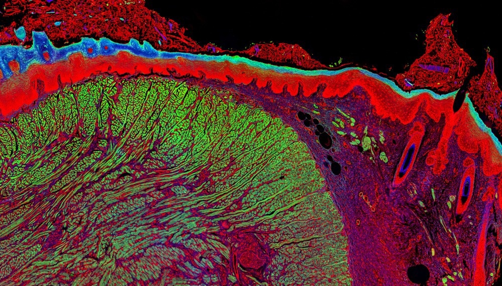
A fluorescence sensor is a device that emits a fluorescent signal when interacting with a target molecule, ion or biological feature.1 Here, we discuss the applications of fluorescence sensors to cellular biology.

Image Credit: Caleb Foster/Shutterstock.com
What is Fluorescence Imaging?
There are many different kinds of fluorescence sensors available. For example, the sensor can be a fluorescent molecule, a nanoparticle or a composite.
An ideal fluorescence sensor will have a high quantum yield of emission, have a high degree of specificity for their desired target, and emit at an easily detectable wavelength.2 Depending on the design of the fluorescence sensor, the sensor may only fluoresce on interaction with the target species.
Another common sensor design is the use of photochromic systems that undergo a wavelength shift, which is the emission wavelength upon recognition of the substrate of interest.3 Many of these types of fluorescence sensors are based on photoswitchable chemical species and can be useful with methods such as super-resolution microscopy for cell imaging.4
For the life sciences, fluorescence sensors have been an invaluable tool in illuminating the secrets of cells. By designing fluorescence sensors sensitive to different aspects of the cell environments, including substrate, pH or local forces, fluorescence sensors have made imaging of specific cell organelles straightforward.5
As each organelle has different characteristic features, specific fluorescence sensors with characteristic emission colors can differentiate and visualize specific cell regions.
For example, fluorescence sensors, combined with the higher imaging resolution capabilities offered by microscopy methods like super-resolution microscopy, have been used to image the amyloid fibrils not just when they are fully formed but during their formation process.4
The team hopes their insights into the amyloid formation process will help provide further insights into how to treat diseases such as Alzheimer's and Parkinson’s through disruption of the formation of plaques that are thought to contribute to the disease advancement.
Cell Imaging
High-resolution imaging of cells is a critical tool in many different areas of the life sciences.6 High throughput cell imaging is often used as a tool for drug design and discovery, as well as a way of understanding the mechanisms underpinning certain diseases for targeted therapeutic development.
Cell imaging studies are also an important part of toxicology and safety assessment studies of new drugs and therapies and are often a regulatory requirement.
Developments in the types of species used as fluorescence sensors have made live cell imaging of different cell structures possible.6 This means that dynamical processes, such as how a cell interacts with a therapeutic species or how proteins fold, deform and undergo turnover, can be monitored in real-time.
With an ever-growing variety of photo-controllable and photo-activeable fluorescence sensors available, it has become possible to tag and image specific parts of protein structures and also measure protein kinetics.
Many diseases specifically affect protein folding and structures. As a result, imaging methods that can retrieve structural and dynamic information on these processes are highly valuable in understanding fundamental structural biology and therapeutic development.
Many research teams are using genetically modified fluorescent proteins as fluorescent sensors that can be incorporated into biological species such as plants to monitor complex biological processes such as metabolism.7 The fluorescent sensors and their spatially localized emission can be monitored with microscopy to track how specific metabolites are transported through the plant structure and be used to identify where particular metabolites are collected or processed.
Toxicology studies also make use of similar approaches with fluorescent probes to monitor the metabolism of potentially harmful chemical species and which regions of the cell a potential toxin can interact with.8 This can be an efficient way of determining the potential risks of a new chemical species with unknown toxicity and screening for potential damage mechanisms.

Image Credit: Micha Weber/Shutterstock.com
Future Developments
One challenge with biological imaging is the issue of poor transparency in the optical regime of many biological tissues. For performing in vivo measurements, which would be necessary for live point-of-care measurements in patients, optical fluorophores are not suitable. The exception is if the detection of the fluorescence can be performed deep in the tissue.
A highly active area of research in fluorescence sensors is the development of sensor species that emit in the near-infrared biological windows.9,10 Not only does this allow for better transmission of the emitted light for better quality images with reduced acquisition times, but opens up a range of possibilities for deep tissue imaging and reduces the required sample preparation to prepare sufficiently thin tissue slices.
Fluorescent sensors are also becoming increasingly common in medical devices.11
There is a constant influx of new fluorescence sensors being developed, further enhancing the types of biological structures and chemical species that can be detected. A greater variety of fluorophores is also aiding the imaging techniques that can be used for gaining greater insight into the structure and behavior of cells.
A wide variety of fluorescent sensor compounds are now available, including antibodies that are compatible with advanced microscopy methods. This is accompanied by a large number of shared protocols for the necessary cell treatments to use said fluorescent probes, as well as specifically designed imaging protocols for commercial instrumentation.
This often includes the use of specific filters for spectral imaging studies that can help distinguish between different cell organelles.
Sources:
Demchenko, A. P. (2008). Introduction to fluorescence sensing. Springer Science & Business Media. doi.org/10.1007/978-1-4020-9003-5
Basabe-desmonts, L., et al. (2007). Design of fluorescent materials for chemical sensing. Chemical Society Reviews, 36, pp. 993–1017. doi.org/10.1039/b609548h
Qin, M., et al. (2015). Photochromic sensors : a versatile approach for recognition and discrimination. Journal of Materials Chemistry C, 3, p. 9265. doi.org/10.1039/c5tc01939g
Kaur, A., et al. (2022). A Fluorescent Sensor for Quantitative Super-Resolution Imaging of Amyloid Fibril Assembly. Angewandte Chemie, 61, e202112832. doi.org/10.1002/anie.202112832
Zhu, H., et al. (2016). Fluorescent Probes for Sensing and Imaging within Specific Cellular Organelles. Accounts of Chemical Research, 49, pp. 2115–2126. doi.org/10.1021/acs.accounts.6b00292
Specht, E. A., et al. (2017). A Critical and Comparative Review of Fluorescent Tools for Live-Cell Imaging. Annual Reviews of Physiology, 79, pp. 93–117. doi.org/10.1146/annurev-physiol-022516-034055
Sadoine, M., et al. (2021). Designs , applications , and limitations of genetically encoded fluorescent sensors to explore plant biology. Plant Physiology, 187, pp. 485–503. doi.org/10.1093/plphys/kiab353
Moczko, E., et al. (2016). Fluorescence-based assay as a new screening tool for toxic chemicals. Nature Publishing Group, 6, p. 33922. doi.org/10.1038/srep33922
Feng, Z., et al. (2021). Perfecting and extending the near-infrared imaging window. Light: Science & Applications. doi.org/10.1038/s41377-021-00628-0
Zhao, M., et al. (2021). Chemical Science Activatable fluorescence sensors for in vivo bio- detection in the second near-infrared window. Chemical Science, 12, pp. 3448–3459. doi.org/10.1039/D0SC04789A
Gunnlaugsson, T., et al. (2017). Fluorescent chemosensors: the past, present and future. Chemical Society Reviews, 46, p. 7105. doi.org/10.1039/c7cs00240h