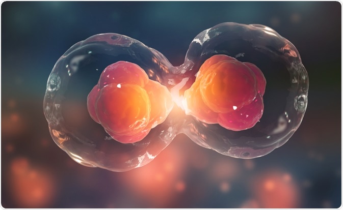
Eukaryotic cells pass through distinct phases known as the cell cycle. The phases of the cycle allow the cell to replicate its genetic material and to divide and produce two identical daughter cells. Two gap phases, known as G1 and G2, an S phase (or synthesis) phase where genetic material is replicated, and a final M (for mitosis) phase where the cell divides and the replicated material is split between the resultant identical daughter cells, are all the phases that make up the cell cycle. Here, we will discuss each phase in detail, and consider what happens when the cell cycle is disturbed.

Cell Division. Image Credit: Yurchanka Siarhei/Shutterstock.com
Gap phase 1
During gap phase 1, or, G1, cells increase in size, and their cellular contents are duplicated, apart from their chromosomes. During this phase, metabolic changes occur to prepare the cell for division. At the point known as the restriction point, the cell becomes committed to division, and the cycle moves onto the S phase.
Synthesis phase
The synthesis phase defines the stage where the cell’s genetic material is replicated. Each of the cell’s 46 chromosomes is duplicated, resulting in each chromosome developing into two sister chromatids.
Gap phase 2
During gap phase 2, known also as G2, the cell grows more and its organelles and proteins develop in anticipation of cell division. Metabolic changes occur at this stage to help the cell assemble cytoplasmic materials in preparation the cell for mitosis and cytokinesis.
The first three phases of the cell cycle, G1, S, and G2 are collectively known as interphase. The following phase of the cell cycle, mitosis, is divided into the five distinct stages of prophase, prometaphase, metaphase, anaphase, and telophase. We describe the processes involved in each stage of mitosis below.
Mitosis
Mitosis is a form of cell division that produces two identical daughter cells containing the same genetic material as the parent cell. During mitosis, the chromosomes produced during the preceding synthesis phase are divided so that each of the resultant daughter cells contains one copy of every chromosome.
The first stage of mitosis is prophase. Roughly half of the time that cells spend in mitosis is spent in prophase. During this stage, the cell’s nuclear membrane is degraded, causing small vesicles to form and the nucleus to disintegrate. Next, the centrosome is replicated to create two daughter centrosomes. These structures then move towards opposing ends of the cell. It is the responsibility of the centrosomes to organize the synthesis of the microtubules that come together to create the mitotic spindle. At the end of prophase, the chromosomes condense and can be seen to contain two sister chromatids that are joined by a region of DNA known as the centromere.
Prometaphase follows prophase. During this second stage of mitosis, the centromeres of the chromosomes are led by their centromeres to the equatorial plane in the center of the cell and settle in the region known as the metaphase plate. Here, the fibers of the mitotic spindle bind to a structure located on each side of the centromere known as the kinetochore. During this phase, the chromosomes continue to condense.
Next, the cells enter metaphase, where the chromosomes simply align themselves along the metaphase plate, leaving the two chromosomes side by side along the central horizontal plane of the cell.
Once the cell’s chromosomes have aligned, the cell enters anaphase. Cells spend an estimated 3% of their total time in mitosis in anaphase. During this stage, the cell’s centromeres divide, and each pair of chromosomes is pulled apart. Each half of the chromosome moves away from its previously adjoining half as the spindle fibers pull them towards opposite ends of the cell. These separated sister chromatids are referred to as daughter chromosomes.
Now the cell is ready to enter telophase. This is the shortest and final phase of mitosis. During telophase, many of the processes that occurred in prophase are reversed. This phase sees the reformation of the nuclear membrane, enclosing the chromosomes at either pole of the cell. Following this, the chromosomes then uncoil and become diffuse. At this stage, the spindle fibers are no longer visible.
Following this final stage of mitosis is cytokinesis where the final step of cell division takes place. This results in the formation of two identical daughter cells. From here, the cell then reenters interphase, before it begins the process of replication again.
Mitosis and disease
The rapid and uncontrolled proliferation of cells is the hallmark of cancer. Mitosis, as discussed above, is the process that governs cell replication. For this reason, the process of cell division has become a key focus of cancer research.
Over recent years, evidence from scientific studies has helped to expand our knowledge of the genes that are implicated in mitosis, and how malfunctions during mitosis can influence the establishment and growth of cancer in humans. Further research into mitosis may, therefore, contribute to the development of more effective therapeutics for a range of cancers.
Sources:
- Collins, K., Jacks, T. and Pavletich, N., 1997. The cell cycle and cancer. Proceedings of the National Academy of Sciences, 94(7), pp.2776-2778. https://www.pnas.org/content/94/7/2776.short
- Kastan, M. and Bartek, J., 2004. Cell-cycle checkpoints and cancer. Nature, 432(7015), pp.316-323. https://www.nature.com/articles/nature03097
- Schafer, K., 1998. The Cell Cycle: A Review. Veterinary Pathology, 35(6), pp.461-478. https://journals.sagepub.com/doi/abs/10.1177/030098589803500601
Further Reading
Last Updated: Feb 26, 2021