Glycans play important roles in various biological functions. Increasingly, research is showing that they are implicated in disease. To understand the role of glycans in the body, mass spectrometry has emerged as the primary tool for documenting these biological structures. Here, we discuss why investigating glycans is important and how mass spectrometry is used to do this.

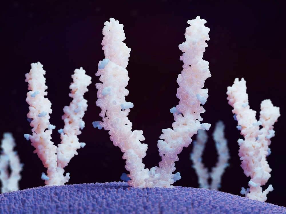
Image Credit: Juan Gaertner/Shutterstock.com
The Importance of Studying Glycans
Glycobiology is fundamental to understanding the workings of biological mechanisms. The discipline refers to the study of the structure, function, and biology of carbohydrates, known as glycans. Complex and diverse glycans exist in all living cells and appear essential to all life forms, making their study crucial to understanding life.
We know that glycans play a crucial role in various biological processes. Therefore, this also means that glycan activity is also associated with disease. In recent years, it has become increasingly clear that glycans are implicated in a wide range of diseases. Insight into changes in glycan activity that underly disease may be vital to developing novel therapeutic interventions.
Studies have shown that many diseases are related to circulating antibodies directed against specific glycan molecules. Evidence shows that Alzheimer’s disease, various forms of cancer, pulmonary diseases, schizophrenia, autism, cardiovascular disease, and others are associated with glycan activity.
For example, glycans have emerged as important mediators and moderators of cancer cell mechanotransduction. Increased glycan content has been observed in cancer cells that form stiffer tumors.
Glycans are also associated with mental health. For example, a glycan affixed to neurexin was recently discovered essential to its function in the central nervous system. When absent, it may increase the risk for schizophrenia.
Illuminating the secret world of your glycans
Therefore, to understand disease, both physical and mental, we must study glycans to understand their role, which may potentially lead to the development of new therapeutics.
Leveraging Mass Spectrometry Imaging in Glycobiology
Mass spectrometry has been successfully used to capture spatial distributions of a range of biomolecules, including proteins, lipids, metabolites, and glycans. It is considered a tool that is fundamental to accelerating medical development. Recently, it has emerged as the go-to tool for investigating glycans. Various studies have used mass spectrometry to investigate glycans and their role in various biological systems, including host/pathogen and host/commensal interactions, mammalian reproduction, and glycosylation.
In simple terms, mass spectrometry is the production and detection of ions that are categorized by their mass-to-charge ratios. As a result of this detection, a mass spectrum is produced, which plots the relative abundance of ions as a function of the mass-to-charge ratio.

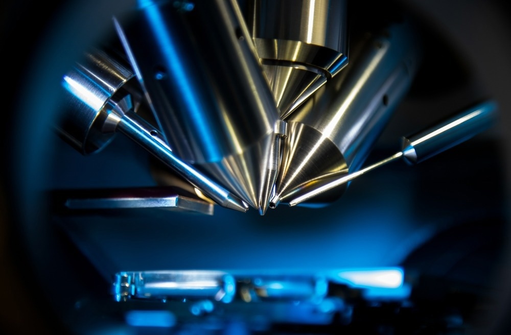
Image Credit: Intothelight Photography/Shutterstock.com
Matrix-Assisted Laser Desorption/Ionization (MALDI) and Electrospray Ionization (ESI) are currently the two most commonly used mass spectrometry techniques for analyzing glycans. Both techniques detect free glycans as a metal adduct in a positive ion mode and as anion-adducted or deprotonated species in a negative ion mode. Developments in mass spectrometry have helped to push glycomics studies forward.
Mass spectrometry imaging, a development of the mass spectrometry technique, has also emerged as a prominent tool in glycobiology. Mass spectrometry imaging is now the primary tool for studying glycans in the brain, partly because it has overcome some of the challenges traditional histostaining methods face. Mass spectrometry imaging does not require information regarding the analyte it is to study, unlike immunohistochemistry, making the technique particularly useful for studies of discovery.
The technique of mass spectrometry imaging is unique in that it combines histology together with mass spectrometry. This allows for the label-free detection of vast amounts of compounds in a single sample without the need for additional methods such as extraction, purification, or separation. Also, mass spectrometry imaging can incorporate quantitative techniques that allow for direct quantitative imaging.
The most commonly used mass spectrometry imaging techniques used to visualize the glycol include matrix-assisted laser desorption ionization-mass spectrometry imaging (MALDI-MSI), desorption electrospray ionization mass spectrometry imaging (DESI-MSI), and secondary ion mass spectrometry (SIMS).
Future Directions for Mass Spectrometry in Glycobiology
There is the potential to develop mass spectrometry tools to help further accelerate glycobiology research. For example, there is an opportunity to develop mass spectrometry imaging techniques to generate 3D representations of glycans within an organ or tissue. The recent combination of ion mass spectrometry with other imaging methods has bridged the gap between non-invasive functional imaging and fundamental biology.
In the future, we can expect developments of 3D mass spectrometry techniques to continue to bridge this gap and particularly help advance glycobiology research in the brain. Much work will be needed before the technique is fully developed, however.
Overall, the development of mass spectrometry in glycobiology will likely lead to deepening our understanding of the pathology of several diseases, which may help establish novel therapeutic approaches.
Sources:
- Han, L. and Costello, C.E. (2013) “Mass spectrometry of Glycans,” Biochemistry (Moscow), 78(7), pp. 710–720. Available at: https://doi.org/10.1134/s0006297913070031.
- Hasan, M.M. et al. (2021) “Mass spectrometry imaging for glycome in the brain,” Frontiers in Neuroanatomy, 15. Available at: https://doi.org/10.3389/fnana.2021.711955.
- Klionsky, Y. and Antonelli, M. (2020) “Thyroid disease in lupus: An updated review,” ACR Open Rheumatology, 2(2), pp. 74–78. Available at: https://doi.org/10.1002/acr2.11105.
- Mealer, R.G. et al. (2020) “Glycobiology and schizophrenia: A biological hypothesis emerging from Genomic Research,” Molecular Psychiatry, 25(12), pp. 3129–3139. Available at: https://doi.org/10.1038/s41380-020-0753-1.
- Purushothaman, A., Mohajeri, M. and Lele, T.P. (2023) “The role of glycans in the mechanobiology of cancer,” Journal of Biological Chemistry, 299(3), p. 102935. Available at: https://doi.org/10.1016/j.jbc.2023.102935.
- Varki, A. (2016) “Biological roles of Glycans,” Glycobiology, 27(1), pp. 3–49. Available at: https://doi.org/10.1093/glycob/cww086.