While the term might sound negative, cell death plays a central role in life, contributing to developing multicellular organisms by depleting dispensable cells and helping eliminate damaged or dangerous ones.
Cell death is a critical process for fighting infections and is associated with many diseases. Therefore, there is a lot of interest in understanding and modulating cell death pathways. Apoptosis and necrosis are the most well-known types, but other cell death pathways have been identified over the years and are attracting increasing interest.
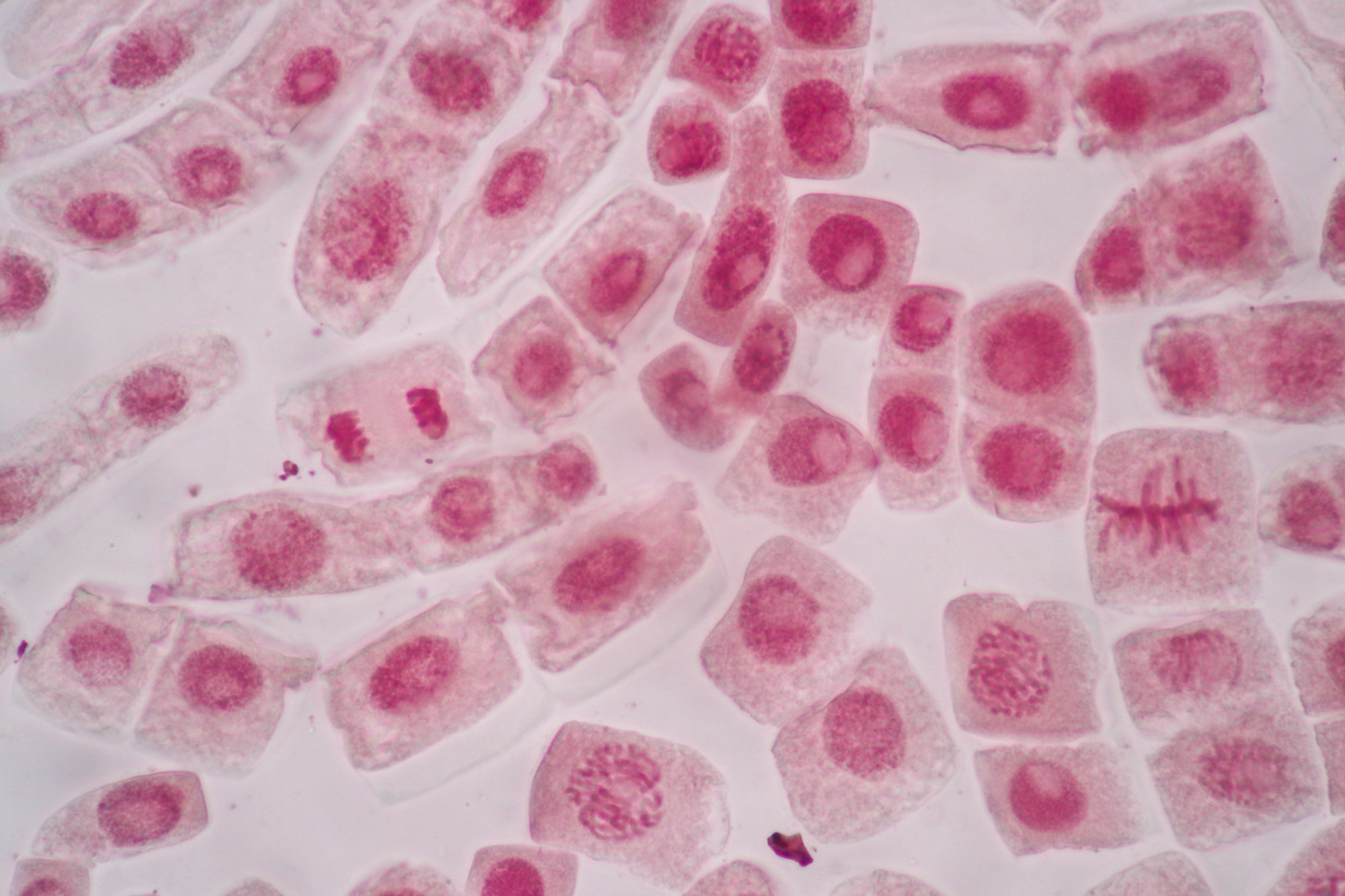
 Image Credit: Rattiya Thongdumhyu/Shutterstock.com
Apoptosis
Apoptosis, sometimes referred to as “cell suicide,” is a programmed mechanism that contributes to cell turnover and the functioning of the immune system, as well as being involved in inflammatory pathologies.
During apoptosis, cells undergo changes such as shrinkage (with reduction in size and loss of water) and DNA fragmentation, where the cell's DNA is cleaved into small fragments. These changes lead to the formation of apoptotic bodies, which are subsequently recognized and engulfed by surrounding cells and phagocytes.1
There are two major mechanisms for apoptosis – the intrinsic and extrinsic pathways. At the heart of apoptosis are enzymes called caspases (a family of cysteine proteases) activated in response to specific signals. Caspases are key regulators of programmed cell death, either playing a role in transmitting cell death stimulus or in its execution.2
Necrosis
Unlike apoptosis, necrosis is an uncontrolled cell death, typically in response to severe injury or infection. While it is a clean, controlled process that maintains tissue health, necrosis can trigger an inflammatory response in the body, leading to further tissue damage.
Necrosis is morphologically characterized by the swelling of cytoplasm and organelles and the disruption of the plasma membrane. This causes loss of integrity and function, ultimately releasing cellular components and cell lysis.
When the cell membrane ruptures, its contents are spilled into the extracellular space, often triggering an inflammatory response. As well as being linked to chronic inflammation, necrosis could also enhance the proliferation of tumors.3
What is Necrosis vs What is Apoptosis?
Beyond Apoptosis and Necrosis: The Spectrum of Cell Death
Although apoptosis and necrosis are the most studied forms of cell death, other types, such as necroptosis and pyroptosis, are also known.
With morphological changes similar to necrosis, necroptosis is a caspase‐independent form of necrotic cell death that results in the rupture of the cell. This process is regulated by specific signaling pathways, and it is often used when apoptosis is inhibited.
Pyroptosis is an inflammatory form of cell death typically triggered by infections. It occurs when activated caspases cleave gasdermins – a family of pore-forming effector proteins – forming pores in the plasma membrane. Pyroptosis is involved in many hereditary diseases, autoinflammatory disorders, and cancer.
Over the last decades, various forms of cell death have been identified, each relying on a different subset of proteins to activate and execute the respective pathway.
These include autophagy‐dependent cell death (ADCD), mitochondrial permeability transition pore (MPTP)‐mediated necrosis, NETosis, and ferroptosis. These forms of cell death add to the complexity of the cell death spectrum and highlight the diversity of the cell’s responses to different stressors or stimuli.1
Subscribe to our Cell Biology Newsletter
Unlocking the Secrets: Studying Cell Death Pathways
The dysregulation of cell death pathways can lead to major disorders, ranging from infection, inflammation, neurodegenerative diseases, and cancer. For instance, the resistance of apoptosis stimulates the multiplication of aberrant cells, eventually leading to tumorigenesis.3
These pathways are attractive targets for therapeutic intervention. In recent decades, the molecular level's understanding of cell death mechanisms has advanced significantly, leading to novel approaches for treating conditions associated with abnormalities in cell death.4
Methods such as the annexin V binding assay and the lactate dehydrogenase (LDH) assay can detect an alteration in the permeability and morphology of the cell membrane.
Apoptotic cells can be identified and quantified based on the dislocation of phosphatidylserine with fluorescently labeled annexin V. LDH appears outside the cell when the cell membrane permeability is compromised.5
The LDH assay is both colorimetric and fluorometric. In the colorimetric assay, a tetrazolium salt is used to detect the cytotoxicity of a compound.
The amount of color produced is measured at 492 nm and is proportional to the number of damaged cells. Similarly, in the fluorometric LDH assay, the light absorbed is directly proportional to cells with compromised membrane permeability.
Methods such as the APO ssDNA assay and TUNEL can detect DNA fragmentation during programmed cell death. The first uses antibodies produced against single-stranded DNA (ssDNA) to find damaged DNA. Instead, the TUNEL assay can quantify apoptotic and necroptotic cells using fluorescence cytometry and microscopy upon detecting labeled DNA strand breaks (DSBs).
Morphological changes, such as cell shrinking, membrane blebbing, and increased cytoplasmic density, can be observed under light microscopy with routine staining methods.
However, light microscopy requires expertise and is low-reproducibility. Conversely, electron microscopy allows the observation of fine ultrastructural changes such as mitochondrial swelling and chromatin condensation (which is visible by light microscopy only at later stages).
Conclusion
Programmed cell death via apoptosis, unregulated necrosis, and other emerging pathways highlight the complexity of cell death.
Several methods exist for measuring and studying cell death-related parameters, each with different specificities, sensitivity, and limitations.
Understanding the different pathways enhances the knowledge of cellular processes and paves the way for innovative treatments for various diseases.
References
- Kist, M. & Vucic, D. (2021). Cell death pathways: intricate connections and disease implications. Embo j, 40, e106700.10.15252/embj.2020106700.
- Yuan, J. & Ofengeim, D. (2024). A guide to cell death pathways. Nature Reviews Molecular Cell Biology, 25, 379-395.10.1038/s41580-023-00689-6. Available: https://doi.org/10.1038/s41580-023-00689-6
- Jan, R. & Chaudhry, G. E. (2019). Understanding Apoptosis and Apoptotic Pathways Targeted Cancer Therapeutics. Adv Pharm Bull, 9, 205-218.10.15171/apb.2019.024. https://www.ncbi.nlm.nih.gov/pmc/articles/PMC6664112/
- Wiman, K. G. & Zhivotovsky, B. (2017). Understanding cell cycle and cell death regulation provides novel weapons against human diseases. Journal of Internal Medicine, 281, 483-495.https://doi.org/10.1111/joim.12609. Available: https://onlinelibrary.wiley.com/doi/abs/10.1111/joim.12609
- Kari, S., Subramanian, K., Altomonte, I. A., Murugesan, A., Yli-Harja, O. & Kandhavelu, M. (2022). Programmed cell death detection methods: a systematic review and a categorical comparison. Apoptosis, 27, 482-508.10.1007/s10495-022-01735-y. https://www.ncbi.nlm.nih.gov/pmc/articles/PMC9308588/
Further Reading
Last Updated: Jun 14, 2024