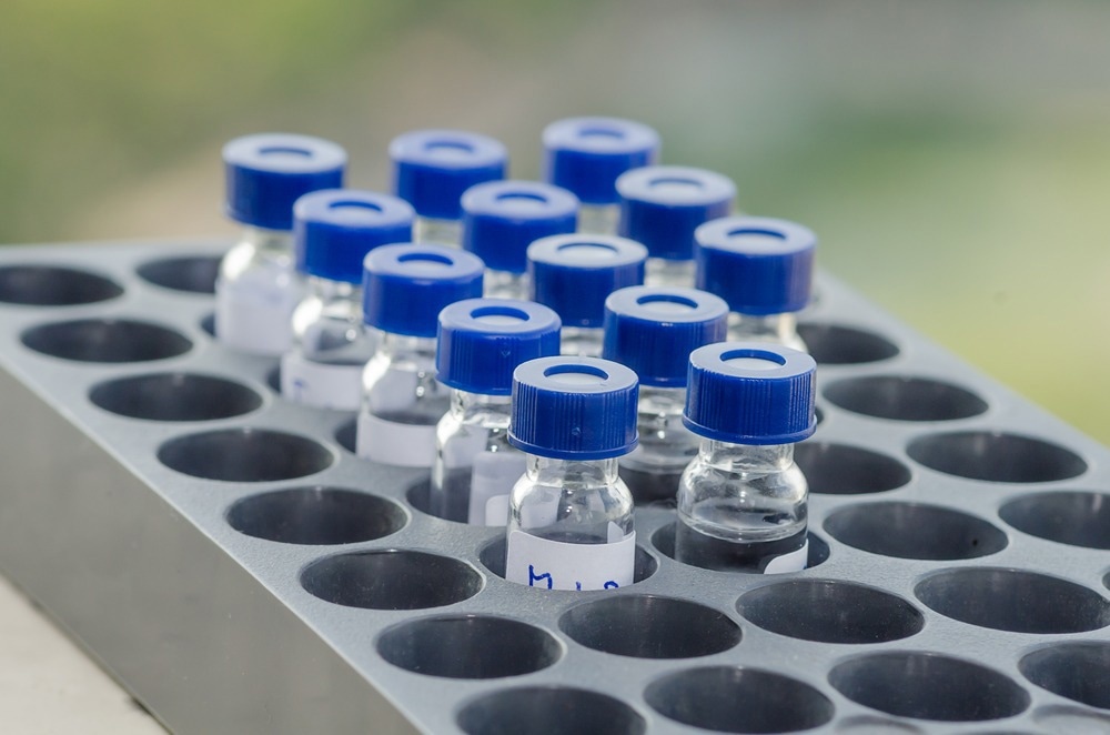
In this article, AZoLifeSciences explores how mass spectrometry can be used to locate proteins in cells.

Image Credit: Rueangwit/Shutterstock.com
Why Is Locating Proteins in Cells Important?
Studying protein localization is vital to cell biology because the location of a protein can reveal the physiological context for protein function. Within eukaryotic cells, membrane-encapsulated compartments are characterized by specific sets of proteins. Each compartment has distinct cellular processes based on these proteins. The location of these proteins within the compartments is essential to providing context for the function of the cell.
Mutations in genes that contribute to the pathology of diseases can alter the process whereby proteins are moved to a specific location in the subcellular environment. Changes to the location of a protein can lead to profound changes in cellular function. Therefore, abnormal protein localization is associated with certain diseases.
Studying protein location helps us understand the pathogenesis of many human diseases, including cardiovascular, cancer, metabolic, and neurodegenerative diseases. Research into protein localization can not only develop our understanding of the underlying mechanisms of disease, but also presents an interesting opportunity to create novel strategies for therapeutic intervention.
What Is Mass Spectrometry?
Mass spectrometry was developed at the turn of the 20th century by JJ Thompson, who used it to uncover the first evidence of nonradioactive isotopes. Mass spectrometry is now used in a wide range of applications across diverse sectors of science.
The analytical tool is used to obtain the mass-to-charge ratio (m/z) of a molecule or molecules present in a sample. The exact molecular weight of the sample can then also be calculated based on this information. Mass spectrometry is often used to identify and quantify unknown compounds as well as determine the structure and chemical properties of molecules within samples.
The process of mass spectrometry involves three stages. In the first, molecules are transformed into gas-phase ions by one of several appropriate techniques, such as nanoelectrospray ionization, so that they can be manipulated and moved via electric and magnetic fields. The resultant ions can be negatively or positively charged, depending on the needs of the experiment.
In the next stage, a mass analyzer is used to sort and separate the ions created in the initial phase. Many mass analyzers are available to conduct this task, and the selection of the appropriate mass analyzer often depends on the requirements of the experiment.
In the final stage, the separated ions are measured and data on their m/z ratios and relative abundance are sent to a computer system which plots this data as a mass spectrum - depicting the m/z ratios against their intensities. Peaks in the mass spectrum represent a unique component in the m/z sample, and the peak’s heights relate to the relative abundance of the component in the sample.
Using Mass Spectrometry to Locate Proteins in Cells
Mass spectrometry is a valuable tool that has significantly advanced our knowledge of cellular composition. Since the early 2000s, systems of organellar proteomics have been available; however, it was not until 2016 that scientists published a method allowing for global dynamic mapping of protein subcellular localization.
A rapid proteomic profiling workflow was developed by a team at the Max Planck Institute of Biochemistry, Germany. The method proved to be a powerful tool, which the scientist used to produce an extensive database of human protein subcellular localizations and organellar composition.
The study created organellar maps through fractionation profiling. The team used HeLa cells - “immortal” cells that were discovered in 1951 and are now often the human cell line of choice in biomedical research. The cells were labeled metabolically and mechanically lysed to induce the release of the cell’s organelles. Next, light labeled lysate was subjected to centrifugation. The heavy labeled lysate was also centrifuged to generate the organellar pellet. Finally, the heavy organellar ‘reference’ fraction was added to equal protein amounts of the five light membrane sub-fractions. At this point, each of the sub-fractions was analyzed by mass spectrometry to generate SILAC ratios for each.
These ratios were then converted to enrichment over reference and profiles were generated by plotting the median values of organellar marker proteins. Heavy-labeled nuclear, organellar and cytosolic fractions were also subjected to mass spectrometry, which revealed the global distribution of proteins across these fractions.
The method was successful in determining protein subcellular localization and it has been developed in the years that have followed since its inception.
Future Directions
The use of mass spectrometry in protein localization is still relatively new. As research continues, it is likely that this method will be further adapted so that its applications can be expanded. The use of mass spectrometry in protein localization stands to further our knowledge of the pathology of numerous diseases. It also has the potential to suggest novel therapeutic approaches by highlighting biological mechanisms to target. In the future, mass spectrometry will likely become increasingly important in this field.
References and Further Reading
Itzhak, D.N. et al. (2016) Global, quantitative and dynamic mapping of protein subcellular localization, eLife, 5. https://elifesciences.org/articles/16950
Laurila, K. and Vihinen, M. (2009) Prediction of disease-related mutations affecting protein localization, BMC Genomics, 10(1), p. 122. https://bmcgenomics.biomedcentral.com/articles/10.1186/1471-2164-10-122.
Luheshi, L.M., Crowther, D.C. and Dobson, C.M. (2008) Protein misfolding and disease: From the test tube to the organism, Current Opinion in Chemical Biology, 12(1), pp. 25–31. https://www.sciencedirect.com/science/article/abs/pii/S1367593108000392.
Park, S. et al. (2011) Protein localization as a principal feature of the etiology and comorbidity of genetic diseases, Molecular Systems Biology, 7(1). https://pubmed.ncbi.nlm.nih.gov/21613983/.
Last Updated: Jun 14, 2023