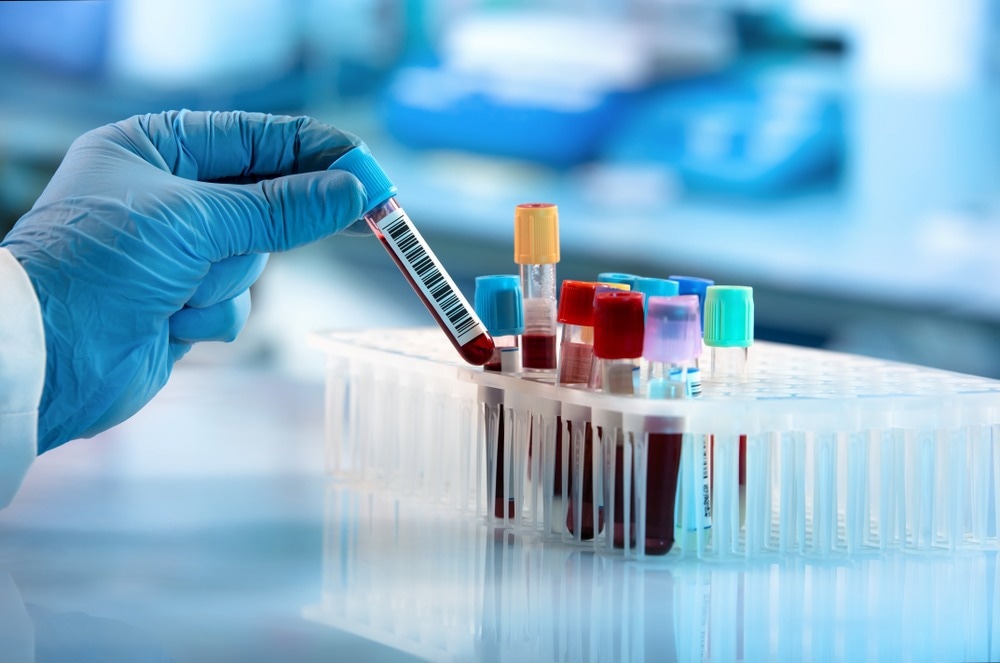
In this article, we look at new research investigating how Raman spectroscopy could be used to find bacteria in blood samples.

Image Credit: angellodeco/Shutterstock.com
Bacterial Infections are a Leading Cause of Death Worldwide
Bacterial infections impact countries across the globe, taking over 6.7 million lives each year and accounting for 8.7% of annual healthcare spending.
Typically, individuals diagnosed with a bacterial infection are given a set of broad-spectrum antibiotics as treatment. However, these antibiotics have contributed to an alarming rise in antibiotic-resistant bacteria. Furthermore, some of the methods used to diagnose these bacterial infections are associated with time-consuming procedures and high costs.
Raman spectroscopy, often in conjunction with other technologies like artificial intelligence (AI), has been proposed as a way to speed up the diagnostic process, monitor patients and recommend appropriate treatments.
What is Raman Spectroscopy?
Raman spectroscopy is used to study the molecular structure of a sample; it relies on a monochromatic light source (such as lasers) to generate photons from the sample. These photons scatter at the same and different energies at each chemical bond within the sample structure. The device then detects the changes and translates into graphs or “spectra”. Each molecule has a characteristic vibrational pattern known as its own unique spectral “fingerprint”.
Raman spectroscopy has moved out from the confines of materials science and emerged as a method for the rapid and minimally invasive identification of bacterial infections, bacterial cells, and antibiotic resistance without using labels to destroy the sample. Recently, it has been able to detect samples without the need for cultures.
Bacteria can be Detected in Blood Using Raman Spectroscopy
Analysis of bacterial samples can provide information on chemical composition and biomolecular structures. DNA, RNA, proteins, lipids and carbohydrates can be detected – often called the whole-organism fingerprint. Since every cell species and strain has a unique molecular structure, they generate a unique spectral fingerprint used for their identification.
Traditionally, Raman scattering efficiency is generally low – subtle spectral differences can easily be masked by background noise, requiring a high signal-to-noise ratio for high identification accuracy. Unfortunately, this can increase measurement times and prohibit high-throughput single-cell techniques. In addition, large numbers of clinically relevant spectral patterns from species and strains of interest require comprehensive datasets that are not readily available and may not be gathered in studies that focus on differentiating between species, strains, or antibiotic susceptibilities in bacteria.
Using AI with Raman Spectroscopy
In a study by Ho et al. (2019) published in Nature, the application of AI with Raman spectroscopy in the identification of bacteria in samples was explored by training a convolutional neural network (CNN) to classify noisy bacterial spectra by strain (or isolate in this study), empiric treatment and antibiotic resistance. The CNN was trained using a reference dataset of 60,000 spectra from 30 bacterial and yeast isolates, covering bacterial infections representative of most infections found in intensive care units worldwide, the data was augmented with 12,000 spectra from clinical patient isolates.
From a 30-class isolate identification task, the CNN outputs a probability distribution across 30 reference isolates and the maximum is taken as the predicted class. The test occurs on an independent test dataset gathered from separately cultured samples. Overall, the accuracy is around 82.2%, with key observations showing that gram-negative bacteria are primarily misclassified as other gram-negative bacteria.
Although the same is generally true for gram-positive bacteria, the misclassification occurs within the same genus. This can be taken further with the CNN recommending empiric treatment based on the bacterial species; the CNN will arrange the 30 isolates into groupings based on the recommended empiric treatment, and the average accuracy was around 97%.
Extension of the CNN to new clinical settings could require additional training and continuous evaluation. The study achieved signal-to-noise ratios that are lower than typical reported bacterial spectra while still achieving a comparable or improved identification accuracy on more isolate classes than typical Raman bacterial studies.
A Novel Approach Using AI with Raman Spectroscopy
An approach by Safir et al. (2023) involved the development of a culture-free analysis method using an acoustic bioprinter to digitize samples into millions of droplets containing a variety of cells. The cells were then identified with SERS and machine learning. Samples of mouse red blood cells were printed and suspended in a solution with spike-ins of gram-positive Staphylococcus epidermidis, gram-negative Escherichia coli and gold nanorods (GNRs) for signal enhancement.
The Raman spectra of each printed droplet were collected and used to train machine-learning algorithms; training involved using uniform cell-type spectra as well as mixed-cell samples to identify the droplet constituents.
Overall, the cellular classification accuracies were ≥99% from uniform cell samples and ≥87% from mixed-cell samples, with validation from electron microscopy of the samples to confirm the presence of specific cells.
Signals were enhanced significantly with the use of GNRs by between 300-1500x compared to controls, tackling issues with signal-to-noise ratios. Using acoustic-printing-based droplet SERS in conjunction with AI allows for rapid digitization of cells from fluid samples in picoliter droplets with minimal sample contamination.
The applications for this method of cell identification are promising; using acoustic droplet ejection-based SERS could enable culture-free cellular identification and monitoring from samples with low concentrations or samples with species that are difficult to culture. Remarkably, the method is low cost as well as rapid, reliable and low contamination, which paves the way for their application in low-cost point-of-care diagnostics.
Conclusions
Raman spectroscopy is a remarkable tool that is amplified when used with techniques such as CNN and AI. Low-cost, rapid and reliable methods of identification are much needed to optimize the clinical diagnostic and treatment process with results produced in less time, with lower cost and appropriate treatment thus limiting antimicrobial resistance and improving patient outcomes.
Sources:
Safir, F., et al. (2023). Combining Acoustic Bioprinting with AI-Assisted Raman Spectroscopy for High-Throughput Identification of Bacteria in Blood. Nano Letters, 23(6), pp.2065–2073. doi.org/10.1021/acs.nanolett.2c03015.
Lunter, D. J., et al. (2022). Novel aspects of Raman spectroscopy in skin research. Experimental Dermatology, [online] 31(9), pp.1311–1329. doi.org/10.1111/exd.14645.
Wang, L., et al. (2021). Applications of Raman Spectroscopy in Bacterial Infections: Principles, Advantages, and Shortcomings. Frontiers in Microbiology, [online] 12, p.683580. doi.org/10.3389/fmicb.2021.683580.
Rebrosova, K., et al. (2022). Raman Spectroscopy—A Novel Method for Identification and Characterization of Microbes on a Single-Cell Level in Clinical Settings. Frontiers in Cellular and Infection Microbiology, 12. doi.org/10.3389/fcimb.2022.866463.
Last Updated: Oct 20, 2023