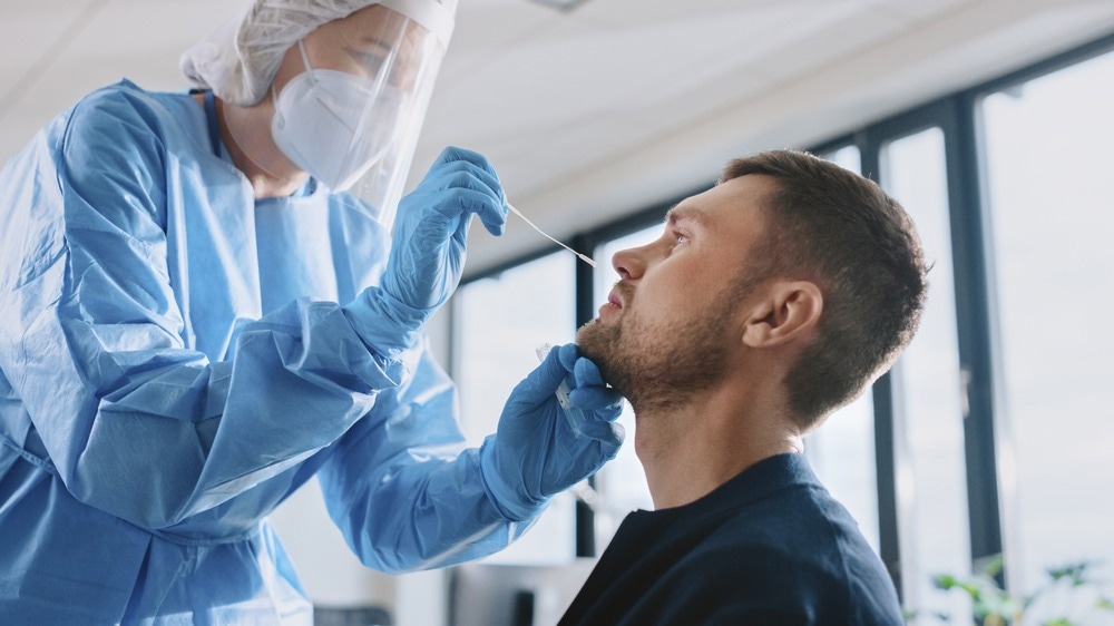
The coronavirus disease 2019 (COVID-19) pandemic led to the global use of polymerase chain reaction (PCR) testing, a moniker that has become commonplace. Though PCR delivers testing results with adequate veracity, at an appropriate turnover rate, faster and larger-scale testing should be implemented, which will be instrumental in combating this pathogen.

Image Credit: Gorodenkoff/Shutterstock.com
For the sake of countries and continents with limited resources, rapid & reagent-free spectroscopic screening has been implemented to detect SARS-CoV-2. Spectroscopy, in this case, is an umbrella term, and scientists are now faced with the question of which spectroscopic methodology is superlative of its counterparts. The modus that will be covered in this report are Raman Spectroscopy (RS), Attenuated Total Reflection Fourier Transform Infrared (ATR-FTIR), and Proton Nuclear Magnetic Resonance (1H-NMR).
Spectroscopy as a Science
Research in this field is dependent on the efforts of data processing, AI (machine learning), and microbiology and should reflect the standards and practices of these concrete fields. When performing spectroscopy, note that its accuracy relies on the quality of samples, the protocols in use, software tuning, and hardware configurations. Many papers advocating for this technique impress the use of nasopharyngeal samples collected via swabs or serum. If nasopharyngeal samples are chosen, they should be inserted into sterile tubes harboring 1-3 mL of a viral transport media.
Compared to the typical PCR testing modus, the benefits of this technique are the quicker detection times, the lesser extent of personnel training, and the circumvention of using probes and primers, which saves on kits and resources.
Raman Spectroscopy (RS)
An often-preferred method when diagnosing ailments at the point of care, RS has the ability to identify many different biomarkers, among them are RNA, antigen, and IgM and IgG antibodies. This technique can relay a snapshot of data regarding the composition of a certain biological species. This can be done through the testing of blood, urine, saliva, etc.
If several sample methods are available, the researcher in question will often opt for serum (80-100 μL depending on the assay) for spectra reading in triplicate. A wavelength of excitation must be chosen, a wattage of laser power must be selected, and collection time should be specified to achieve the appropriate spectral range. Ana Cristina Castro Goulart et al. published a paper depicting their optic measurements when analyzing human serum transfected with COVID-19. They excite within the 830nm range with a 350mW laser, achieving a spectral resolution of 4cm-1 between the spectral range of 400cm-1 and 1800cm-1. Collections of times were spaced at 30 seconds.

Attenuated Total Reflection Fourier Transform Infrared (ATR-FTIR)
FTIR encompasses a 97.8% accuracy, in addition to limiting post RNA extraction times from hours to minutes. It incorporates machine learning and is label-free and simple in its application. Used in other afflictions such as diabetes, cancer, and hypertension, FTIR assays the vibrational optical nature of bonds to determine what is in each sample. An IR Spectrum has a transmittance percentage (light recorded by the detector) on the Y-Axis and a wavenumber on the X-Axis. Some common regions of interest within the IR spectrum are the alkyne C-H stretch (3300 cm-1), the OH stretch (3200-3400cm-1) for hydroxyl and carboxylic acid functional groups, and alkenyl C-H stretches (3000cm-1).
The identifying biomarkers that make up the COVID-19 capsid and viral load are easy to identify and are, in turn, well suited for this device. In hypercytokinemic (severe immune reaction via too many cytokines released into the blood) cases of COVID-19, an N-H deformation stretch will appear at peaks around 1299 (94/95/96) cm-1. Contracting COVID-19 leads to a sharp increase in proteins generated, triggering a hyperinflammatory response that could lead to organ dysfunction. A direct consequence of this is using phosphates as a modular block for energy, indicated by a band at 1146cm-1.
These signals can even provide crucial information on whether certain medicinal moieties are functioning when given to the patient. A peak at 2838 cm-1 is characteristic of a methoxy functional group, which perfectly relates back to esters used in heavy medications like anesthetics and other analgesics.
Proton Nuclear Magnetic Resonance (1 H-NMR) in COVID testing
Rather than using a Raman spectroscopy stretching spectra, the H-NMR chemical shifts can reveal diagnostic markers of the COVID-19 infection by combined diffusional and relaxation editing (DIRE). This concept of photoconversion explores the biochemistry of human plasma and can associate changes in metabolic biomarkers like glycoproteins, amino acids, and lipids. These can be discerned through NMR fit to mass spectrometry.
DIRE characterizes the translational mobility of compounds; useful when identifying smaller biomarkers that bind to larger proteins. Small molecules diffuse very fast in a sample, though their diffusion reduces when they are bound to proteins. By taking note of the change in diffusion, we can characterize the binding of COVID metabolites and smaller substructures other than spike or envelope proteins. This is why DIRE is widely used and is one of the standard approaches used in drug discovery. Samantha Lodge et al. identified the main diagnostic markers for SARS-CoV-2 using DIRE, finding N-acetylglycoprotein peaks GlycA and GlycB with signals at δ 3.20–3.30.
Sources:
- Gurbanov R, Tunçer S, Mingu S, Severcan F, Gozen AG. Methylation, sugar puckering and Z-form status of DNA from a heavy metal-acclimated freshwater Gordonia sp. J Photochem Photobiol B. 2019 Sep;198:111580. doi: 10.1016/j.jphotobiol.2019.111580. Epub 2019 Jul 31. PMID: 31394353.
- Lodge, S., Nitschke, P., Kimhofer, T., Wist, J., Bong, S. H., Loo, R. L., Masuda, R., Begum, S., Richards, T., Lindon, J. C., Bermel, W., Reinsperger, T., Schaefer, H., Spraul, M., Holmes, E., & Nicholson, J. K. (2021). Diffusion and Relaxation Edited Proton NMR Spectroscopy of Plasma Reveals a High-Fidelity Supramolecular Biomarker Signature of SARS-CoV-2 Infection. Analytical chemistry, 93(8), 3976–3986. https://doi.org/10.1021/acs.analchem.0c04952
- Khan, R. S., & Rehman, I. U. (2020). Spectroscopy as a tool for detection and monitoring of Coronavirus (COVID-19). Expert review of molecular diagnostics, 20(7), 647–649. https://doi.org/10.1080/14737159.2020.1766968
- Kitane, D.L., Loukman, S., Marchoudi, N. et al. A simple and fast spectroscopy-based technique for Covid-19 diagnosis. Sci Rep 11, 16740 (2021). https://doi.org/10.1038/s41598-021-95568-5
- Nogueira, M. S., Leal, L. B., Marcarini, W. D., Pimentel, R. L., Muller, M., Vassallo, P. F., Campos, L., Dos Santos, L., Luiz, W. B., Mill, J. G., Barauna, V. G., & de Carvalho, L. (2021). Rapid diagnosis of COVID-19 using FT-IR ATR spectroscopy and machine learning. Scientific reports, 11(1), 15409. https://doi.org/10.1038/s41598-021-93511-2
- Goulart, A., Silveira, L., Jr, Carvalho, H. C., Dorta, C. B., Pacheco, M., & Zângaro, R. A. (2022). Diagnosing COVID-19 in human serum using Raman spectroscopy. Lasers in medical science, 1–10. Advance online publication. https://doi.org/10.1007/s10103-021-03488-7
- Yang, B., et al. Sub-nanometre resolution in single-molecule photoluminescence imaging. Nature Photonics. doi.org/10.1038/s41566-020-0677-y.
Further Reading