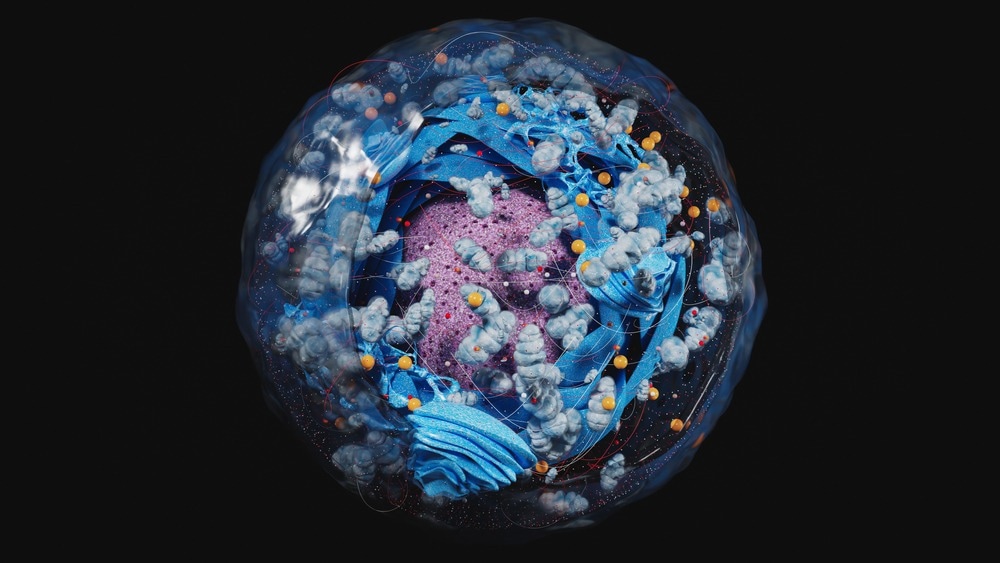
For survival, every cell requires energy. This energy source is produced within a cellular structural component called the mitochondrion (plural form: mitochondria) in the form of adenosine tri-phosphate (ATP).

Image Credit: CI Photos/Shutterstock.com
As well as energy production, mitochondria have many other functions, which include the production of reactive oxygen species (ROS), modulation of calcium, steroid synthesis, nucleotide metabolic processing, intermediary metabolism regulation, and apoptosis. Thus, mitochondria are vital structures for cell survival and therefore play an important role in every cell in our body, particularly important for cells with a high demand in energy, such as the eye cells.
Mitochondria and the Eye
Found abundantly in retina photoreceptors, such as cones, mitochondria are situated within the inner segment of the cone, more specifically, the ellipsoid that lies proximal to the outer segment (OS), which is known to be light-sensitive. With this understanding of mitochondrial location within photoreceptor cones, it is assumed that its primary function is to provide an abundant supply of ATP to facilitate the phototransduction process, that is, to convert light energy in the form of photons to electric membrane potential change across a cell.
However, though, recent research indicated that the ATP that photoreceptors of the retina utilize is primarily from glycolysis, an energy-producing process that occurs in the cytosol of the cell, and thus independent from mitochondria. This would suggest that the high number of mitochondria found within photoreceptor cones may not be there for its ATP-producing capacity.
This, therefore, begs the question, what is the function of abundant mitochondria found in photoreceptor cones?
The answer may lie in how other cellular structural components could affect light reaching the light-sensitive OS. It is understood that evolutionary pressure has evolved the retina to detect light favorably, thus managing the path of light to reach the OS for its detection.
Evidence of this is shown in the photoreceptor cones in nocturnal mammals, whereby the tight arrangement of chromatin within the nuclei is suspected to reduce the scatter of light, therefore focusing light more for more precise vision, which is essential in dark environments due to limited light.
Further evidence of retina evolutionary adaptation is from the Müller glial cells, support cells found in the retina, which have been noted to guide light for intraretinal light scatter reduction, thus facilitating enhanced light detection.
Hence, it is probable that the abundance of retinal mitochondria assists light in reaching the OS, augmenting light detection and improving vision. Interestingly, oil droplets are observed proximally to the OS, that is, in the distal cone IS, and due to the high refraction index of oil droplets, it is thought to focus light on the OS for detection. Likewise, the lipid membrane of mitochondria also has a high refraction index, indicating that mitochondria may have a similar optical role; however, their internal structures, such as the cristae, may perturb the concentration of light.

Image Credit: Africa Studio/Shutterstock.com
Mitochondria having Optical Functions
To test whether mitochondria bundles found in the retina could have a role beyond energy production, that is, to have optical functions, a team of researchers led by Wei Li et al. (2022) went and studied the mitochondrial bundles of photoreceptor cones found within the retina of squirrel, with both live imaging and computer simulations.
The research team managed to isolate photoreceptor cone mitochondria from the ground squirrel retina and then shone light through the bundle of mitochondria contained in the membrane. The observation via confocal microscopy was that after the light passed through the bundle of mitochondria, the resulting light emerged as a bright, concentrated beam. This finding provides evidence that the mitochondria have optical functions within the eye, which may be essential to energy production. It seems that the mitochondria may be assisting in the focusing light for its detection by the light sensitive OS of the retina, and in turn, providing a clearer and more precise vision.
Further evidence of the mitochondria's optical functioning is provided by the computer-simulated comparison of light passing through differing shaped bundles of mitochondria. During the ground squirrel's active state, the mitochondria bundle is organized and elongated, whereby the light emerging from this structure was seen to be well-focused. In contrast, the bundle of mitochondria in a hibernating ground squirrel, with its disorganized and compressed structure, did not nearly focus light as well.
Conclusion
Li and the team have paved the way for a new understanding of mitochondria function and will provide an avenue for further research into eye diseases in which mitochondria are implicated. In addition, it provides a truly remarkable insight into how organelles and structural components of the cell may have other functions unknown to us.
Sources:
- Ball J, Chen S, Li W. Mitochondria in cone photoreceptors act as microlenses to enhance photon delivery and confer directional sensitivity to light. Science Advances. 2022;8(9).
- Kanow M, Giarmarco M, Jankowski C, Tsantilas K, Engel A, Du J et al. Biochemical adaptations of the retina and retinal pigment epithelium support a metabolic ecosystem in the vertebrate eye. eLife. 2017;6.
- Solovei I, Kreysing M, Lanctôt C, Kösem S, Peichl L, Cremer T et al. Nuclear Architecture of Rod Photoreceptor Cells Adapts to Vision in Mammalian Evolution. Cell. 2009;137(2):356-368.
- Agte S, Junek S, Matthias S, Ulbricht E, Erdmann I, Wurm A et al. Müller Glial Cell-Provided Cellular Light Guidance through the Vital Guinea-Pig Retina. Biophysical Journal. 2011;101(11):2611-2619.
Further Reading