Autopsy and Virtopsy
A part of forensic medicine is to elucidate the scientific medical findings that can be found in deceased individuals.
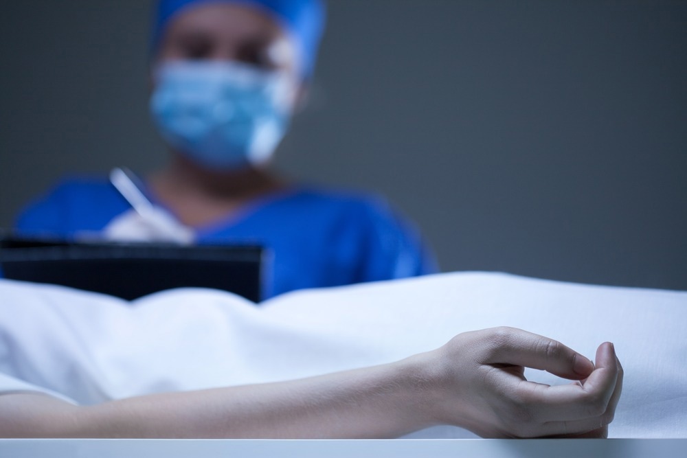
This endeavor can be approached through autopsy, where the whole surface of the body, along with its cavities, is examined to document all findings that could be relevant to a forensic case. Autopsies are considered highly unpleasant for the general public due to their invasive nature. However, they provide crucial information that can connect the deceased and what or who killed them, ultimately developing a forensic reconstruction of what exactly transpired.
Nevertheless, considering the deceased person's relatives is also of utmost importance, as they may find traditional autopsies disrespectful and appalling. Therefore, the concept of virtopsy was introduced, which is meant to be an unbiased analysis of physical features and evidence depicted in a three-dimensional way.
'Virtual autopsies', are they the way of the future?
Early Attempts and On-going Progress
In the late 1970s, computed tomography (CT) was used for a gunshot pattern injury. However, the attempt did not gain much popularity due to the low image quality and resolution. In the late 1980s, Kalender et al. introduced spiral CT as a gateway to 3D data acquisition. Despite this breakthrough, the interest of forensic scientists wasn't sparked.
In 2000, the University of Bern, thanks to "The virtopsy project" introduced a scalpel-free autopsy that was made possible by using modern medical imaging. Specifically, four cornerstone techniques are employed in a virtopsy: 3D surface scanning, computer-aided design photogrammetry, multi-slice computed tomography, and magnetic resonance imaging. The aforementioned techniques provide a complete 3D view of the body without destroying the geometry, which is always the case with traditional autopsies.
Virtopsy in Detail
Virtopsy requires the preparation of the body to be examined by placing small disks along its surface, which facilitates the alignment of the surface scans with the interior scans. In some devices, this step can be performed automatically. After setting up the disks, stereoscopic cameras capture a 3D color image of the body. Upon completion of the image creation, the investigators can manipulate and examine the image.
Following the surface scan, the body is wrapped up in blue bags, which allow the X-rays to pass, and then the body is laid onto the CT/MRI table. CT and MRI scans happen in quick succession. By taking advantage of the X-rays properties to be absorbed differently by each tissue type, a 3D visualization is produced with different colors for each type of tissue that has been scanned. The result of all the scans can be displayed on a large touch-sensitive LCD screen where the pathologist can peel through layers of virtual skin.
What are the Advantages and Disadvantages of Virtopsy?
The advantages of a virtopsy extend further than being a scalpel-free, ethical evolution of conventional autopsy. There is no hazard of infections to the health care workers since no incisions are required and, therefore, no blood. It is a less time-consuming method compared to its non-digital counterpart, allowing the corpse to be released right after scanning.
Another merit of virtopsy is that it provides an efficient way to match a probable weapon by studying wounds without altering the body structure. Specifically, in a case of a bullet injury, the entry wound, the tract of the bullet in the body, and the exit wound, can be easily assessed. There are, of course, a few downsides when it comes to opting for virtopsy. In some countries, the initial cost for acquiring the equipment may not be feasible.
Besides the cost, the database for comparing wounds, pathological conditions, etc., is still under development and thus very limited. It can also be challenging to comprehend which wounds were inflicted on the deceased ante- or post-mortem; some tissue injuries may not be easily spotted.

Image Credit: ESB Professional/Shutterstock.com
Current Status of Virtopsy and Wider Use
In Japan, post-mortem computed tomography (PMCT) has been widely applied for three major tasks: 1) screening the cause of death, 2) screening bodies for autopsy, and 3) Obtaining supplemental information for autopsy. In a study conducted in Japan, questionnaire sheets were distributed, regarding the use of PMCT, to 183 major medical establishments facilitating emergency rooms.
Out of all medical establishments, 67% responded, and it was found that 89% of the respondents use PMCT. So, in a well-developed country like Japan where the equipment for a virtopsy is not scarce, the technique is becoming a staple.
Virtopsy has been used in various judicial cases, including analysis of the depth of obstruction in a case of demise due to asphyxiation. Precise comparison of ante- and post-mortem records of restorative materials and their variations under extreme temperature, simulating a mass disaster environment. Radiographic examination has been used to identify the pathologic lesions or implants placed in a body part of a mass disaster victim. This can aid in personnel identification by comparing with ante-mortem records. The use of photogrammetry has achieved simulation and analysis of bite marks, lip prints, and fingermark ridge patterns.
General effectiveness of the Method and a Step Forward
The accuracy of virtopsy is well documented and supported by the most up-to-date scientific literature. Virtopsy has about an 80% success rate in identifying the correct cause of death, which is comparable to what would have been identified by a conventional autopsy. It has a high potential for accuracy and can recognize the lines of fractures more precisely and more frequently than an autopsy.
Skull pathologies, primary and secondary trauma details, depth of injury, and small bone injuries are better visualized with virtopsy. Despite its many benefits, virtopsy will become the state-of-the-art technique only when a validated and comprehensive database for injury comparison is established and when the imaging equipment needed becomes more affordable.
Further Reading
Last Updated: Dec 29, 2022