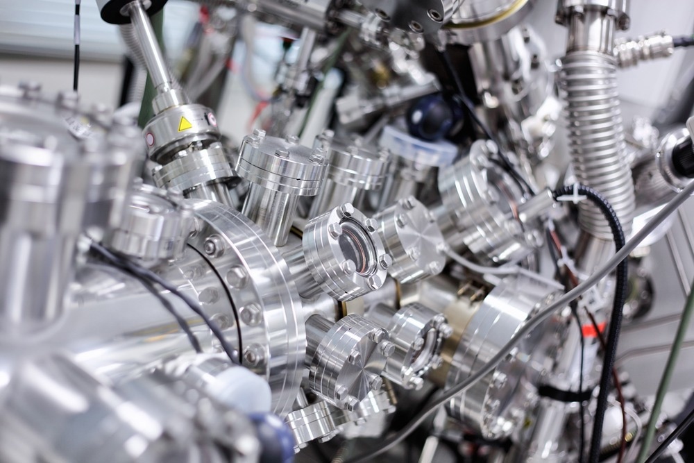
Known as one of the most powerful techniques for surface characterization, X-ray photoelectron spectroscopy (XPS) can identify the elements on the surface of a sample and give information on their chemical bonding.

Image Credit: Alexander Gatsenko/Shutterstock.com
XPS is based on the photoelectric effect, where metal surfaces emit electrons upon light irradiation. Particularly, in an XPS experiment, a sample is bombarded with an X-ray beam, and the kinetic energy of the electrons emitted is measured.
This kinetic energy equals the energy of the incident X-rays (a known parameter) minus the binding energy and the spectrometer work function (a constant value). Therefore, the binding energy of a particular element – independent of the X-ray source – can be calculated, allowing one to determine the sample's composition.
In addition, since the chemical environment affects the binding energy, XPS enables the determination of the neighboring atoms and the oxidation state of the elements. It is possible to distinguish between C-C, C-O, or C-F, based on the electronegativity trend.
Similarly, the oxidation state of most metals can be identified. Atoms that exhibit a positive charge will possess higher binding energies. Concerning titanium's oxidation state, for instance, when moving from Ti(0) to Ti(IV), the binding energy increases from 453.9 eV to 458.7 eV.
In a typical XPS spectrum, the y-axis is the intensity, expressed in counts/s, while the binding energy (in eV) is on the x-axis, with values decreasing from left to right. The energies and intensities of the peaks enable the identification and quantification of the elements on the surface.
The peak area can also be used to obtain the element concentration on the surface since it is a measure of the relative amount of that particular element.
Main Components of XPS Instruments
The main components of an X-ray photoelectron spectrometer are the X-ray source, extraction lenses, analyzer, and detector.
The X-ray source is designed to provide a high fluence of X-rays since the number of electrons emitted is proportional to the X-ray source intensity. Most instruments have Mg and Al sources, with energies of 1253.6 eV and 1486.6 eV, respectively.
The extraction lenses define the acceptance angle for collecting the electrons emitted from the sample and, in some cases, can also enable small spot analysis by controlling the area of the sample from which electrons are collected.
The most common analyzers are concentric hemispherical analyzers consisting of two hemispheres. The electrons enter the analyzer through a slit, and only electrons with a specified energy can travel through.
Finally, the detectors are typically electron multipliers. On average, an experiment can last 30–60 min, depending on the number of high-resolution scans and the concentrations of the elements in the sample.
The experiment takes place in an ultra-high vacuum (UHV) environment, with pressures generally in the range of 10-9-10-10 mbar. The main reason for UHV is the necessity to remove air molecules that can scatter the emitted electrons. Moreover, XPS is very sensitive to surface contamination, and UHV prevents contaminants from sticking to the sample surface.
What are the Strengths and Weaknesses of XPS?
One of the main advantages of the technique is the surface sensitivity and the ability to give information on the sample composition up to a depth of approximately 10 nm. Another key strength is the ability to distinguish differences in chemical environments.
XPS can detect all elements (apart from hydrogen and helium) with detection limits of approximately 0.1–1 atomic percent. It does not require using standards for quantitative determinations and is relatively nondestructive.
On the other hand, XPS instruments are quite expensive, and the analysis time is relatively long. The need for an ultrahigh vacuum is another limitation. Processes involving semi-volatile liquids or porous samples that absorb gases cannot be studied via XPS.
Nevertheless, current research is focused on developing near ambient pressure XPS (NAP-XPS, also called environmental XPS), operating at 25 mbar, and some instruments are already commercially available.

Image Credit: Ico Maker/Shutterstock.com
Applications
Thanks to the ability to identify the chemical functional groups present on the surface of virtually any material, there is an enormous number of applications for XPS, ranging from polymer characterization, analysis of nanomaterials, and bioengineering.
XPS is widely used to characterize a wide range of polymers, such as polyethylene terephthalate (PET), one of the most common polymers used in synthetic fibers and bottle production. The binding energy profiles allow us to identify the C-N, C-O, and C=O bonds.
A variant of XPS called angular resolved X-ray photoelectron spectroscopy (ARXPS) provides the opportunity to examine thin layers and interfaces such as films and coatings.
XPS also plays an important role in nanotechnology since it can detect the presence of elements and their relative concentrations on the surface of nanostructured materials (e.g., nanoparticles). The impurities or contaminants introduced during the synthesis and handling of nanomaterials can also be determined via XPS.
Much research is focused on using XPS in bioengineering applications, particularly in the analysis and characterization of devices or materials. This includes the detection of oxidation on the surface or the segregation of additives and low-molecular-weight fragments.
The presence of contaminants (i.e., silicones), or residual lubricants used during the manufacturing process that may affect either the biological interactions or the function of a material, can also be determined. Surface modification processes can also be optimized following XPS characterization.
Other applications include the adsorption studies of proteins and other biomolecules at interfaces. Thanks to the wide range of elements that can be analyzed and the sampling depth often larger than the dimensions of most adsorbed proteins, both the substrate and the protein can be detected simultaneously.
XPS is, therefore, well-suited for the characterization of biological interactions. It is worth noting that XPS cannot differentiate between proteins, so it is not suitable for studying the adsorption of protein mixtures.
Sources:
- Stevie, F. A. & Donley, C. L. (2020). Introduction to x-ray photoelectron spectroscopy. Journal of Vacuum Science & Technology A, 38, 063204.10.1116/6.0000412
- Engelhard, M. H., Droubay, T. C. & Du, Y. (2017). X-Ray Photoelectron Spectroscopy Applications. Encyclopedia of Spectroscopy and Spectrometry. 3rd edition, 716-724. Oxford:Academic Press. 10.1016/b978-0-12-409547-2.12102-x
- Mcarthur, S. L. (2006). Applications of XPS in bioengineering. Surface and Interface Analysis, 38, 1380-1385.10.1002/sia.2498
Further Reading