The wavelengths of X-ray light has a range in the electromagnetic spectrum from 10 to 0.01 nanometres (nm), where 0.1 nm is a typical wavelength used for crystallography due to it being on the same scale as atomic radii or covalent bonds.

Image Credit: Isuaneye/Shutterstock.com
As such, X-ray crystallography is a technique used not only to understand chemical bonds and non-covalent interactions but also to determine the structure of proteins and biological macromolecules. The ability of X-ray crystallography to elucidate structural information of proteins provides a broad range of applications in the biological sciences.
X-ray crystallography reveals information about atomic radii and thus chemical bonding, which led to discoveries in chemistry about bonding arrangements in molecules and the resonance of various aromatic molecules.
Discoveries of bond lengths led to the finding that chemical bonds are in a state of resonance, which has large implications in the field of chemistry. The strength of a bond and therefore the bond order can be calculated by knowing the distance between two bonded atoms, which is made possible by the use of X-ray crystallography.
Thus, many types of bonds were discovered by this approach (such as double and quadruple metal-metal bonding), as well as research into “back bonding” and supramolecular chemistry being made possible.
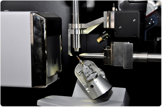
Single-crystal X-ray diffraction measurements involve the mounting of a crystal and then illuminating it with a monochromatic beam of X-rays. This results in a diffraction pattern of spots known as reflections.
These two-dimensional images taken at different orientations of the crystal are converted into a three-dimensional model that shows the electron density within the crystal. This conversion is calculated by Fourier transform mathematical algorithms and combined with chemical information about the sample.
Scattering Techniques of X-ray Crystallography
X-ray crystallography is based on the principle of elastic scattering where the wavelength of the X-ray light does not change whether it is X-rays entering or leaving the crystal. This is important when determining the distribution of atoms and bonds that would generate the largest scattering within the crystal.
Although the single-crystal X-ray diffraction method provides the most detailed structural information compared to other techniques. It requires a large pure crystal, which is sometimes difficult to produce. If single crystals cannot be produced, other scattering techniques can be used but will result in less detailed information.
These techniques include powder diffraction, fiber diffraction, and Small-Angle X-ray Scattering (SAXS). The diffraction patterns produced as a result of the electron density within the crystal can be calculated relatively easily.
Weakly scattered beams pass through the crystal without being subjected to re-scattered waves of X-rays known as secondary scattering, which makes analysis difficult. If a crystal is too thick, then secondary scattering is likely to occur, but since X-rays have a weak interaction with the electrons, it is generally not a problem.
Alternative methods of obtaining diffraction patterns can be achieved using electrons and neutrons compared to using X-rays. An advantage is that they produce much stronger secondary scattering for much thinner crystals in a scale of approximately 100 nm.
This corresponds to the size of some biological systems like viruses and therefore provides applications for macromolecular assemblies, biological molecules, and membrane proteins. Furthermore, the strong scattering effect of electrons allows the elucidation of the atomic structure using small volumes.
Neutron diffraction scatters much more readily from atoms in the crystal so it is very useful for structure determination of light atoms with few electrons, such as hydrogen, which does not produce an X-ray diffraction pattern.
Benefits of a Crystal in X-ray Crystallography
Although atomic structure can be determined from non-crystalline samples, the advantage of producing a single crystal is that they produce a much stronger signal due to their repeating pattern, defined by their unit cells.
The many unit cells of a crystal that are repeated indefinitely in three directions allow for the weak scattering of each unit cell to be concentrated, producing reflections that are constructive interference patterns calculated from the Fourier Transform.
However, if the crystal structure is not pure, but instead disordered or contains more than one species, then it could result in erroneous interpretation. Similarly, liquid or powder samples have disordered molecular structure and limit the structural detail produced in the diffraction pattern.
Applications of X-ray Crystallography
There are broad applications for X-ray crystallography in chemistry, biological science, life science, biochemistry, material science, and mineralogical sciences due to the atomic level resolution and the associated electron distribution.
X-ray, electron, and neutron diffraction all give atomic-level structural information about crystals and non-crystalline material, which can be organic, inorganic, or biological.
This paves the way for many applications, some of which include the identification of antibiotic drugs due to very distinctive X-ray diffraction patterns for each drug.

Image Credit: Sendo Serra/Shutterstock.com
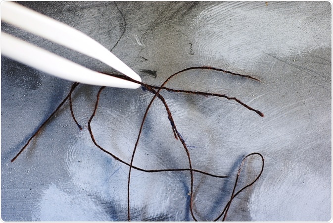
Forensic analysis of fibers and polymers using X-ray diffraction, where it can detect and characterize trace fibers at crime scenes. Furthermore, X-ray diffraction is used for bone analysis to determine the change in bone structure at the atomic level as a result of heating or burning the bones at different temperatures.
References
- Bragg WL; James RW; Bosanquet CH (1921). "The Intensity of Reflexion of X-rays by Rock-Salt. Part II". Phil. Mag. 42 (247): 1.
- Wyckoff RWG; Posnjak E (1921). "The Crystal Structure of Ammonium Chloroplatinate". J. Am. Chem. Soc. 43 (11): 2292.
- Bragg WH (1921). "The structure of organic crystals". Proc. R. Soc. Lond. 34 (1): 33.
- Pauling L (1960). The Nature of the Chemical Bond (3rd ed.). Ithaca, NY: Cornell University Press.
- Powell HM; Ewens RVG (1939). "The crystal structure of iron enneacarbonyl". J. Chem. Soc.: 286.
- Cotton FA, Curtis NF, Harris CB, Johnson BF, Lippard SJ, Mague JT, et al. (September 1964). "Mononuclear and Polynuclear Chemistry of Rhenium (III): Its Pronounced Homophilicity". Science. 145 (3638): 1305–7.
- Wunderlich JA; Mellor DP (1954). "A note on the crystal structure of Zeise's salt". Acta Crystallographica. 7: 130.
- Suryanarayana, C.; Norton, M. Grant (2013-06-29). X-Ray Diffraction: A Practical Approach. Springer Science & Business Media.
- Thangadurai S, Abraham JT, Srivastava AK, Moorthy MN, Shukla SK, Anjaneyulu Y (July 2005). "X-ray powder diffraction patterns for certain beta-lactam, tetracycline and macrolide antibiotic drugs". Analytical Sciences. 21 (7): 833–8.
- Svarcová S, Kocí E, Bezdicka P, Hradil D, Hradilová J (September 2010). "Evaluation of laboratory powder X-ray micro-diffraction for applications in the fields of cultural heritage and forensic science". Analytical and Bioanalytical Chemistry. 398 (2): 1061–76.
- Hiller JC, Thompson TJ, Evison MP, Chamberlain AT, Wess TJ (December 2003). "Bone mineral change during experimental heating: an X-ray scattering investigation". Biomaterials. 24 (28): 5091–7.
Further Reading