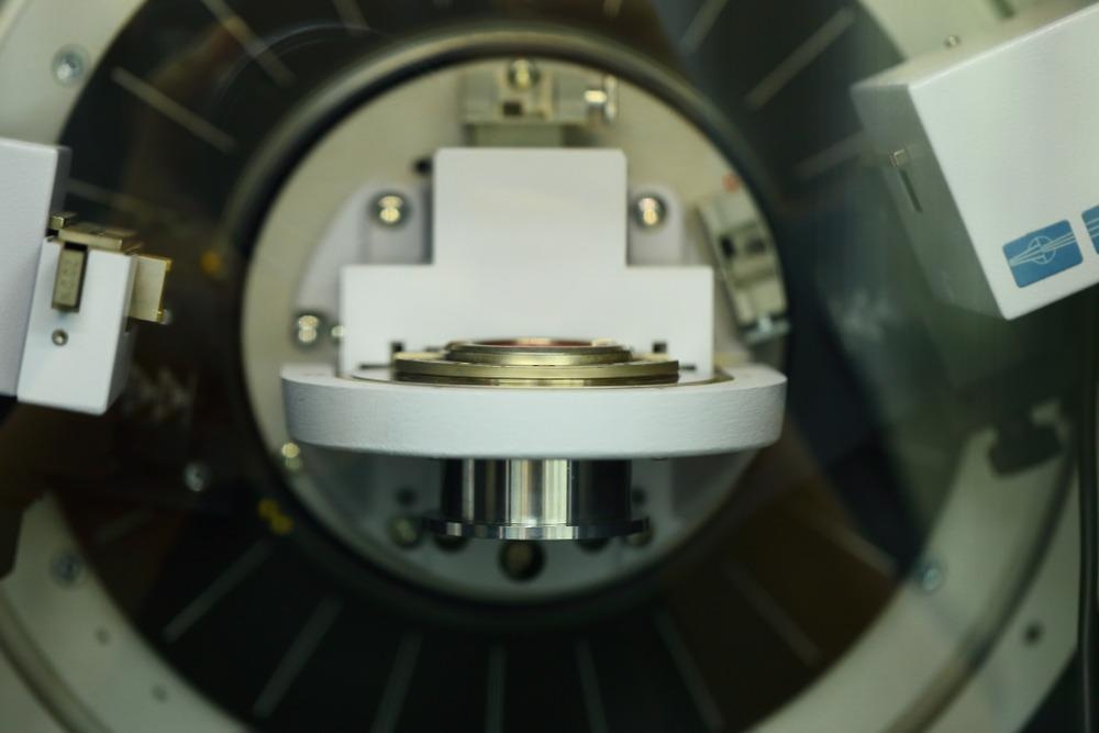
The X-ray diffraction (XRD) technique is used to characterize the structure of a wide variety of materials on the atomic and molecular levels. Such materials are normally crystalline (or partially crystalline) and can range from single crystals, thin films, and mixtures of powders.

Image Credit: AgriTech/Shutterstock.com
XRD is a non-destructive technique and helps scientists to understand how atoms are arranged and how such arrangements can affect the behavior of materials or can be used to determine the structure of complex molecules.
XRD was developed at the beginning of the 20th century and its application to the analysis of crystals led to the award of the Nobel Prize in Physics to Sir William Henry and William Lawrence Bragg in 1915 – one of the many Nobel Prizes associated with XRD.
Principles of XRD – Measuring the Distance Between Atoms
X-rays have wavelengths in the range of 10−10 m, the same order of magnitude as the distance between atoms (measured in Ångströms, Å). In a typical XRD experiment, the sample is illuminated with a beam of X-rays. The X-ray source and the detector move at different angles in a synchronized motion.
Most materials consist of many small crystals. These crystals have a regular arrangement of atoms, each surrounded by a cloud of electrons. When an X-ray encounters an atom, its energy is absorbed by the electrons, and is then released in the form of a new X-ray in a phenomenon called “elastic scattering”.
Scattered waves are subject to interference. When the waves align in what is called “constructive interference” the detected signal is amplified and a reflection is observed. Conversely, in case of destructive interference – signals out of alignment – the signal is destroyed.
Diffraction is the effect of constructive interference and subsequent signal amplification, which takes place at a very specific angle. According to the Bragg equation, a signal is observed when nλ = 2d sinθ.
In this equation n is an integer number, λ (lambda) is the wavelength of the X-ray, d is the distance of the lattice planes for which the peak occurs and θ (theta) is the angle between the lattice planes and the incident beam.
Generally, X-rays are produced by either X-ray tubes or synchrotron radiation. The first is the typical source in a laboratory setting. X-rays are generated by heating a filament to produce electrons, accelerating the electrons toward a target by applying a voltage, and bombarding the target material.
However, nowadays synchrotron facilities are often the preferred sources for X-ray diffraction measurements. Synchrotron radiation is emitted by electrons traveling at near light speed in a circular storage ring. Being up to millions of times more intense than laboratory X-ray tubes, synchrotrons have enabled a wide range of structural investigations and brought advances in numerous fields.
Advantages and Disadvantages
XRD is a very powerful technique. Because of the ability to interact with atoms in crystal structures, it provides extremely useful information on distances and structures on both the atomic and the molecular level, and it is used for identifying unknown minerals and materials.
The high level of structural information that can be obtained is unrivaled by any other technique. The analysis only requires a minimal amount of sample. Several XRD measurement instruments are widely available and data interpretation is relatively straightforward.
However, XRD does have certain limitations. To best identify an unknown material, the sample should be homogeneous. Sample preparation often requires grinding to obtain a powder and typically XRD analysis requires access to standard reference data to make comparisons.
The analysis relies a lot on the availability of high-quality crystals. The preparation of suitable crystals, especially for biological macromolecules like proteins, is a well-known challenge. Nevertheless, advances in the aforementioned synchrotron sources can help overcome this limitation, together with the development of X-ray powder diffraction (XRPD) as an alternative to single-crystal X-ray diffraction (SCXD).
Applications of XRD
The technique is probably the most accurate approach to obtaining detailed structural information for biological macromolecules. The most famous example of XRD applications is probably the discovery of the DNA structure (1953). Using the XRD data produced by Rosalind Franklin, Watson and Crick were in fact able to model the double-helix structure of the molecule of life.
Lately, studies have been conducted on selected virus proteins and also on protein domains that have a critical role in a virus’s lifecycle. Such studies could suggest potential methods for virus inactivation and used XRPD to gather structural information.
Crystallographic studies have been performed on the Dengue virus 3 (DENV3) non-structural protein 5 (NS5), in the methyltransferase domain (MTase), in both the absence and presence of ligands, leading to the identification of potential DENV inhibitors.
XRD is often used to study nanomaterials and obtain information on their morphology, with phase identification being one of the most common applications. An interesting example is the analysis of nanocrystalline ZrO2–Sc2O3 solid solutions – a system of great technological relevance due to its possible application as a solid electrolyte in solid oxide fuel cells.
XRPD measurements were employed to determine the temperature versus composition phase diagram. This revealed that there is a critical crystallite size (approximately 35 nm) above which particular equilibrium phases with low ionic conductivity begin to appear and can have an impact on the material property.
There are also diverse applications in the field of pharmaceutical sciences. It is possible to have information on the composition of drugs, namely the nature and content of active pharmaceutical ingredients (APIs) and excipients in antihistamine tablets (Allegra ®). It is also possible to identify differences in drug composition and XRPD methods have been used for the fast screening of tablets to identify counterfeit medicines.
This is only a limited set of examples. Applications are countless, both in academic research and in the industry (pharmaceuticals, polymers, nanotechnologies, etc.) and despite some limitations, XRD is always the characterization technique of elite.
Sources:
- Stanjek, H. & Häusler, W. (2004). Basics of X-ray Diffraction. Hyperfine Interactions, 154, 107-119.10.1023/B:HYPE.0000032028.60546.38
- Spiliopoulou, M., Valmas, A., Triandafillidis, D.-P., Kosinas, C., Fitch, A., Karavassili, F. & Margiolaki, I. (2020). Applications of X-ray Powder Diffraction in Protein Crystallography and Drug Screening. Crystals, 10, 54.10.3390/cryst10020054
- Abdala, P. M., Craievich, A. F., Fantini, M. C. A., Temperini, M. L. A. & Lamas, D. G. (2009). Metastable Phase Diagram of Nanocrystalline ZrO2−Sc2O3 Solid Solutions. The Journal of Physical Chemistry C, 113, 18661-18666.10.1021/jp904584e
- Thakral, N. K., Zanon, R. L., Kelly, R. C. & Thakral, S. (2018). Applications of Powder X-Ray Diffraction in Small Molecule Pharmaceuticals: Achievements and Aspirations. J Pharm Sci, 107, 2969-2982.10.1016/j.xphs.2018.08.010
Further Reading