Overview
The CELLCYTE X™ from Echo has been specifically developed for live cell imaging and analysis present within the incubator.
Bringing high-throughput live cell imaging to users’ lab
CELLCYTE X™ provides the user the potential to image live cells in real-time from inside the incubator.
From all of such images obtained, the user could completely comprehend and review cell kinetic trends by any chance in time with easy-to-use analysis software
A familiar user experience with intuitive controls
When the company set out to develop the CELLCYTE X™, a look was taken at existing live-cell analyzers and made use of the pain points had by their customers.
From that point, it was known that it was paramount to make a system with simple access to cells and intuitive software to guarantee that the user is frequently in control to successfully execute experiments in a highly efficient manner. The CELLCYTE X provides an experience by giving reliable and reproducible outcomes.
CELLCYTE X™ - Real-time Live Cell Imaging from Inside Your Incubator
Video Credit: Echo
Features—Built for successful experiments
Open design
Simpler maintenance and environmental control
Real-time data analysis
Track live-cell kinetics as they take place
High throughput
Runs up to six vessels concurrently
Versatile
Potential to multiplex experiment with improved contour imaging mode plus 3 FL channels
High data output
Users can capture and store thousands of images for every experiment
Simple setup
Less cords, less issues. Sets up in just minutes
Ease of use
Intuitive UI decreases learning curve and enhances workflow
Improved cell viability
Less disturbances at the time of their experiment decrease cellular abnormality
Applications
CELLCYTE X™ enables the user to answer several questions regarding the cells that may increase during an experiment.
Users can gain a highly comprehensive picture of cellular kinetics via execution of main applications like:
- Confluence
- Reporter genes
- Spheroid growth
- Apoptosis
- Cytotoxicity
- Cell counting
- Transfection efficiency
- Immune cell cluster formations
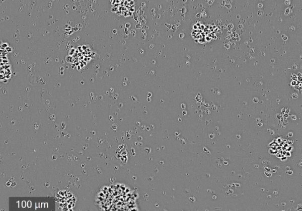
Immune Cell Cluster Formation. Visualize and quantify immune cell interactions and proliferation. Image Credit: Echo
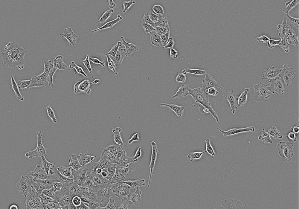
Cell Culture Quality Control. Monitor cell morphology and growth over time from within the incubator. Image Credit: Echo
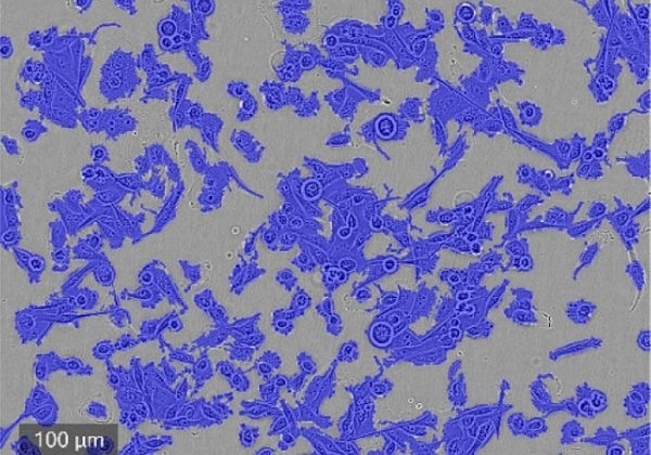
Label-free Cell Confluence. Use label-free segmentation metrics to track cell confluence. Image Credit: Echo
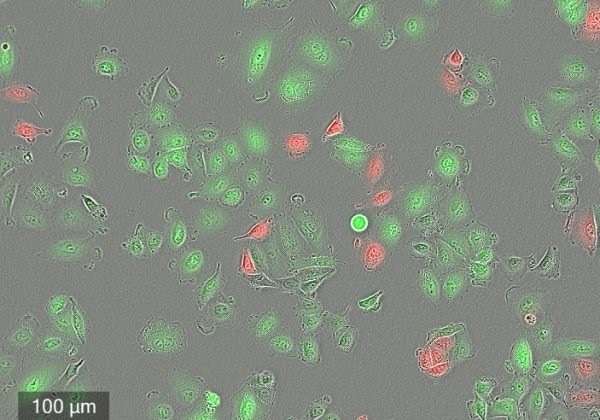
Fluorescence Cell Counting. Measure how live cell populations are growing. Image Credit: Echo
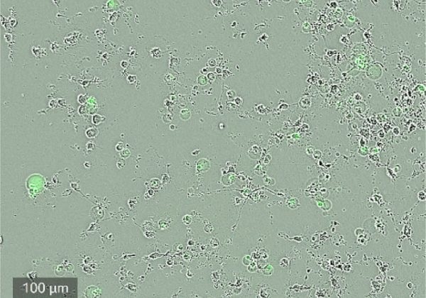
Apoptosis. Monitor cells undergoing cell death by measuring the signal from the activation of caspase-3/7. Image Credit: Echo
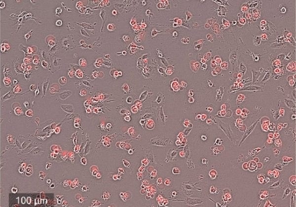
Cytotoxicty. Monitor and quantity the number of cells dying over a period of time. Image Credit: Echo
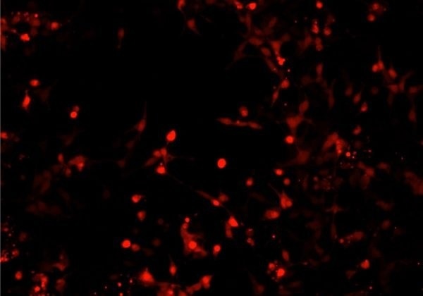
Transfection Efficiency. Monitor the ability of mammalian cells to uptake fluorescently labeled nucleic acids. Image Credit: Echo
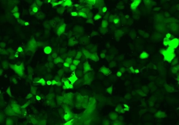
Reporter Genes. Quantify and assess the dynamics of multiple biochemical signals with gene expression or protein activity Image Credit: Echo
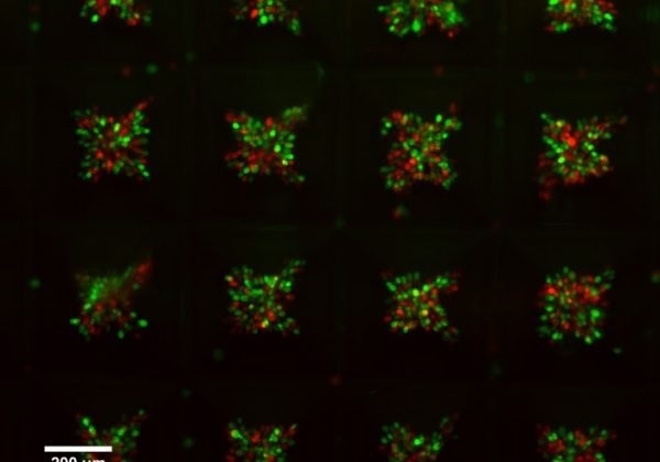
Spheroid Growth. Monitor and quantify single spheroid formation and growth Image Credit: Echo
Integrated data analysis and graphic tools
CELLCYTE X™’s software consists of data analysis and graphing tools to aid the user record high-contrast images and instant visualizations of data points.
Video Credit: Echo