Bruker’s TruLive3D Imager is specifically designed for rapid 3D multi-sample volume imaging of delicate live specimens within their native 3D environments. Employing an optical concept featuring dual-sided illumination and single-lens detection from below (inverted microscope geometry), it achieves fast acquisition speeds, high-resolution imaging, and minimal shadowing effects.
The system's latest technological advancements and optional features, such as the photomanipulation module, make it ideal for time-lapse and high-throughput imaging of 3D spheroids, organoids, 3D cell cultures, and small embryos.
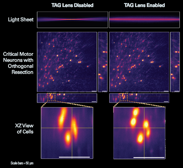
TAG lens disabled generates non-uniform resolution (left). TAG Lens enabled generates a homogeneous illumination profile. Image Credit: Bruker Nano Surfaces and Metrology
Only TrueLive3D Imager Provides:
- Excellent long-term environmental control for live cells and delicate samples
- Elongated sample holders enable the multiplexing of hundreds of samples
- Save time and money by conducting up to six different experimental conditions in a single experiment
- User-friendly mounting with ready-to-go and sterile dishes
Dual-Sided Technology for High-Throughput Discovery
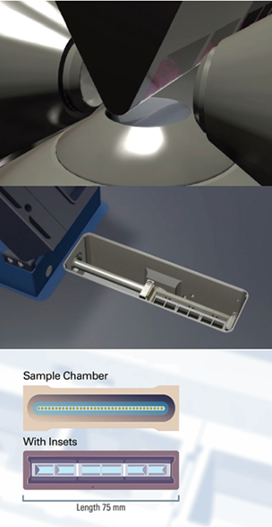
TruLive3D Imager has a large sample holder that is 75 mm long with two sample spaces and three disposable dishes. Image Credit: Bruker Nano Surfaces and Metrology
The TruLive3D Imager is a stable and compact light-sheet solution characterized by dual-sided illumination and single-lens detection from below. This configuration ensures swift acquisition speed, high-resolution imaging, and minimal shadowing effects. The system boasts an impressive resolution down to 255 nm in xy, facilitating the resolution of subcellular structures in living samples with minimal phototoxic effects
The TruLive3D Imager is designed to fit conveniently on a lab bench without the need for an air table, and its piezo-crawler stages ensure precision and longevity, enabling accurate and swift specimen positioning.
Utilizing pseudo-brightfield imaging, samples can be easily located and their health monitored. The system is also optimized for rapid long-term in vivo imaging, offering advanced environmental control for experiments such as spheroid and organoid cell divisions, as well as 3D cell culture growth. Additionally, its large sample holder (75 mm; travel range 66 mm) and TruLive3D Dish series allow for extended imaging under various experimental conditions.
Cutting-edge technologies featuring ultra-fast varifocal lenses ensure precise fine-tuning of the optical system. For adaptive optics, the TruLive 3D Imager utilizes devices driven by an acoustic wave, known as tunable acoustic gradient lenses (TAG lenses). These lenses enhance light-sheet control and uniformity while mitigating axial stretching.
Unique TrueLive3D Imager Features Include
- High-quantum efficiencies of up to 82 % provide excellent image quality
- Simultaneous dual-channel acquisition for high imaging speed
- Stage travel ranges of 66 × 3 × 3 mm for precise sample positioning
- Dual-destriping eliminates strip artifacts
Optional Photomanipulation Module
The TruLive3D Imager can be enhanced with a photomanipulation (PM) module, enabling studies like photoablation, cauterization, optogenetics, and fluorescence recovery after photobleaching (FRAP) with:
- Beam coupled into detection objective
- Diffraction-limited illumination spot
- Flexible region-of-interest (ROI) generation (point, straight line, freeform, circle, and square)
- Laser from continuous wave ultraviolet/visible (CW UV/VIS) to pulsed infrared (IR)
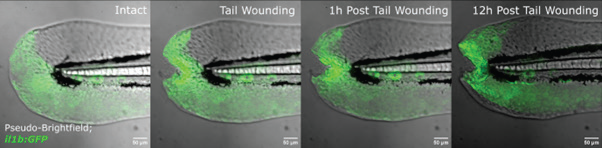
Timelapse of cytokine dynamics in a tail wound assay. Pseudo-Brightfield and il1b:GFP (green) merged. Captured every 5 minutes for 12 hours. Sample courtesy of: Dr. Elizabeth Jerison, Stanford University. Image Credit: Bruker Nano Surfaces and Metrology
TrueLive3D Imager Objective Specifications
Source: Bruker Nano Surfaces and Metrology
| Objective |
Tube lens |
Magnification |
Pixel Size [nm] |
FOV [μm] |
| Nikon CFI APO LWD 25x 1.1 NA |
100 |
12.5x |
520 |
1065 |
| 200 |
25x |
260 |
532 |
| 300 |
37.5x |
173 |
355 |
| 400 |
50x |
130 |
266 |
| Olympus XLUMPLFLN 16x 0.8 NA |
100 |
8x |
813 |
1664 |
| 200 |
16x |
406 |
832 |
| 300 |
24x |
271 |
555 |
| 400 |
32x |
203 |
416 |