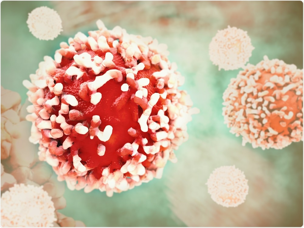
In a genome-wide CRISPR-Cas9 screen published in Nature Immunology in January 2020, a group of researchers described a potential universal target on cancer cells.

Image Credit: Crevis/Shutterstock.com
One of the challenges cancer immunotherapies face is the low recognition rate of cancer cells by T cells. This is due to the high variability of cancer markers such as human leukocyte antigens (HLA) expressed on the surface of cancer cells by different classes of major histocompatibility complex (MHC) molecules, thus making it difficult for T cells to recognize cancerous cells.
A monomorphic MHC-like molecule (MR1) is found to be uniformly expressed on cancer cells and can be targeted by a specific clone of T cells for therapeutic use.
What is MR1?
MR1 is a mammalian cell surface receptor known to present bacterial metabolites to mucosal-associated invariant T cells (MAIT), in a similar way as MHC molecules present peptides. It is evolutionary conserved and monomorphic.
Most studies have shown that MR1 is only expressed when its binding ligand is present; however, there is evidence of basal expression of MR1 on cancer cells.
A new way of targeting cancer cells
In the paper, the group of researchers identified a T cell clone named MC.7.G5 from a healthy donor and found that this T cell population was active against A549 lung cancer cells, although they were HLA-mismatched, and thus MC.7.G5 should not be able to recognize the cancer cells.
This led to the hypothesis that MC.7.G5 can recognize cancer cells via an HLA-independent pathway.
Further experiments show that when MHC molecules were blocked by anti-MHC antibodies, and as a result cancerous molecular marker was not presented on cancer cells by MHCs, MC.7.G5 was still able to recognize the cancer cells.
Moreover, MC.7.G5 was able to kill a panel of established cancer cell lines and primary cancer cells (lung, melanoma, leukemia, colon, breast, prostate, bone and ovarian) even they do not share the same HLA marker.
To be therapeutically relevant, the cancer-killing T cells should not be killing healthy cells. It is found that MC.7.G5 was not toxic to non-cancerous cells, as it did not kill healthy human cells such as smooth muscle, hepatocyte, and lung fibroblast.
Furthermore, to elucidate the specificity of cancer cells targeting, stress was induced in healthy cells by tBHP treatment, exposure to hydrogen peroxide, and irradiation. It is found that MC.7.G5 also did not kill stressed, non-cancerous cells, and sheds light on its specificity in killing cancer cells.
Genome-wide CRISPR-Cas9 screen reveals the target of MC.7.G5
Although MC.7.G5 was able to kill a variety of cancer cells, the target which it recognizes, is unknown. A genome-wide CRISPR-Cas9 screen was performed to knockdown every protein-coding gene in the human genome in a cancerous cell line (HEK293T).
The cancer cells that were protected from killing by MC.7.G5 would give a clue on genes that have important roles in MC.7.G5 recognition of cancer cells. In the genome of the surviving cells, it was found that MR1 was knocked out, hence revealing MR1 as a target.
To further confirm MR1 is a target on cancer cells, the researchers blocked MR1 on the cancer cells by using anti-MR1 antibodies. This abolished MC.7.G5 recognition and killing of cancer cells. This confirmed the role of MR1 in facilitating cancer cell targeting by MC.7.G5.
Moreover, the researchers explored the necessity of MR1 in facilitating cancer cell targeting by MC.7.G5.
Wild-type, MR1 knock out and MR1 overexpression cancer cells were constructed and whether MC.7.G5 kills them or not was investigated. Indeed, MR1 knock out cells were protected against MC.7.G5 and overexpression of MR1 enhanced killing by MC.7.G5, meaning MR1 is necessary for cancer cell targeting.
In vivo experiments
Mouse models are useful for developing new therapeutic targets. Jurkat cancer cells were engrafted into mice and MC.7.G5 was transferred into the mice to test whether the cancer cells can be killed by MC.7.G5 in vivo.
It is revealed that there was a significant decrease in Jurkat cancer cells after 12 days. This shines a light on the in vivo therapeutic potential of MC.7.G5 T cells. To prove MC.7.G5 killing of Jurkat is MR1-dependent, Jurkat cells with MR1 knocked out were also engrafted into the same mice.
It was also found that MC.7.G5 only killed the wild type Jurkat cells, but not the MR knocked out cells. In an in vivo setting, transferal of MC.7.G5 prolonged survival of mice.
More importantly, when the T cell receptor (TCR) of MC.7.G5 was engineered onto the surface of T cells purified from patients with stage IV melanoma by lentiviral transduction, the T cells were able to kill melanomas, establishing the fact that MC.7.G5 was able to redirect patient T cells for cancer immunotherapy.
Future work and implications
This is the first time identifying MR1 as a potential pan-target for cancer immunotherapies and promising results are shown. The next step would be to identify the ligand complexed with MR1 that is recognized by MC.7.G5 and the mechanism of recognition.
Journal reference:
Genome-wide CRISPR–Cas9 screening reveals ubiquitous T cell cancer-targeting via the monomorphic MHC class I-related protein MR1. Nature Immunology, issue 21, 178-185, https://www.nature.com/articles/s41590-019-0578-8#Sec9