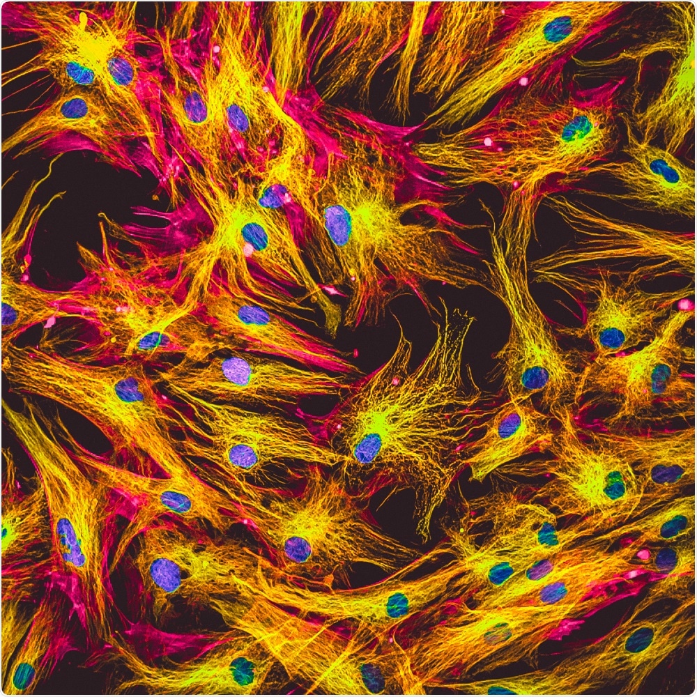
A team of researchers at the University of Illinois published a paper this January in the journal American Chemical Society describing short-wave infrared emitters that were developed for use as molecular labels for microscopy applications, using synthesized semiconductor quantum dots.

Image Credit: Vshivkova/Shutterstock.com
What the team innovated will likely benefit future imaging of blood vessels in deep tissues due to its potential to generate high-contrast and high-resolution images.
Developing SWIR probes for fluorescence microscopy
The use of fluorescent probes has found many uses in biomedical sciences due to its robustness and reliability in the detection, imaging, and tracking of molecules within cells or tissues. Fluorescent probes have been used for many years to identify proteins in samples along with their conformational changes, also, they have been used to determine the location of these proteins in vivo, these functions have allowed fluorescent probes to be used in monitoring biological processes happening in real-time, in vivo.
Scientists have used fluorescent probes to detect human disease, screen populations to determine those at risk, diagnose disease, develop drugs, and evaluate treatment efficacy.
Recent years have seen significant advancements in the use of fluorescent probes, along with the technology and equipment that supports it.
These advancements have helped to rapidly progress methods in microscopic molecular imaging, such as super-resolution optical microscopy and single-molecule imaging that have become well-established in the field of cellular biology.
However, there is one major limitation to the use of fluorescent probes, and this is that cellular components often have an intrinsic fluorescence that can work to obscure the signals emitted by the probes.
Scientists have been working to develop a method to overcome this limitation. Recent research has focused on the development and exploration of nanomaterial-based fluorophores. Researchers are designing and synthesizing these new probes specifically to emit light at wavelengths that span two infrared spectral windows (the first near-infrared window, NIR-I, and the second near-infrared window, NIR-II). The latter of these is known as short-wave infrared (SWIR).
Recent research has seen the rapid expansion of SWIR-emitting nanomaterials. Of these, light-emitting semiconductor quantum dots (QDs) have become frequently used in research because of their advantages, such as broad wavelength tunability, and intense and stable emission that facilitates the tracking of individual molecules.
The team wanted to develop SWIR probes for use in fluorescence microscopy that overcame the limitations of conventional probes.
To do this, they continued to the work on light-emitting semiconductor quantum dots (QDs), creating a new class that produces continuous spectral tenability in the visible, NIR-I, and SWIR ranges.
The research
The focus of the research was to design and develop QDs. The team controlled the emission range by tuning the alloy composition of the QDs, which were constructed of HgxCd1–xSe alloy cores that were red-shifted toward the SWIR via epitaxial deposition of HgxCd1–xS shells.
They demonstrated that the resultant SWIR probes could be created in a consistently small and equal size, allowing for the analysis of their molecular labeling performance over a broad wavelength.
The QDs the team developed was shown to have a quantum yield in the SWIR and a compact size, as well as long-term stability in aqueous media. These characteristics of the synthesized QDs allow for a potentially diverse set of applications of the probes in fluorescence microscopy.
The established SWIR probes showed a significant enhancement of the signal-to-background ratio, meaning that they may be able to be used to create ultrasensitive molecular imaging, capable of detecting low-copy number analytes in biological samples.
Establishment of new SWIR probes
New SWIR probes for fluorescence microscopy were developed by the Illinois-based team. These probes overcome the issue of background fluorescence that currently limits the use of fluorescence microscopy methods.
The SWIR probes yield brightness and stability in aqueous media and can detect analyses present in low concentrations.
Journal references:
Hassan, M. and Klaunberg, B. (2004). Biomedical applications of fluorescence imaging in vivo. Comp Med, 54(6), pp.635-44. https://www.ncbi.nlm.nih.gov/pubmed/15679261
Jensen, E. (2012). Use of Fluorescent Probes: Their Effect on Cell Biology and Limitations. The Anatomical Record: Advances in Integrative Anatomy and Evolutionary Biology, 295(12), pp.2031-2036. https://anatomypubs.onlinelibrary.wiley.com/doi/full/10.1002/ar.22602
Sarkar, S., Le, P., Geng, J., Liu, Y., Han, Z., Zahid, M., Nall, D., Youn, Y., Selvin, P., and Smith, A. (2020). Short-Wave Infrared Quantum Dots with Compact Sizes as Molecular Probes for Fluorescence Microscopy. Journal of the American Chemical Society, 142(7), pp.3449-3462. https://pubs.acs.org/doi/10.1021/jacs.9b11567