Headed by researchers from Japan-based Tokyo University of Agriculture and Technology (TUAT), a group of scientists has effectively established three-dimensional (3D) cultured tissue that imitates liver fibrosis, which is a major trait of non-alcoholic steatohepatitis (NASH).

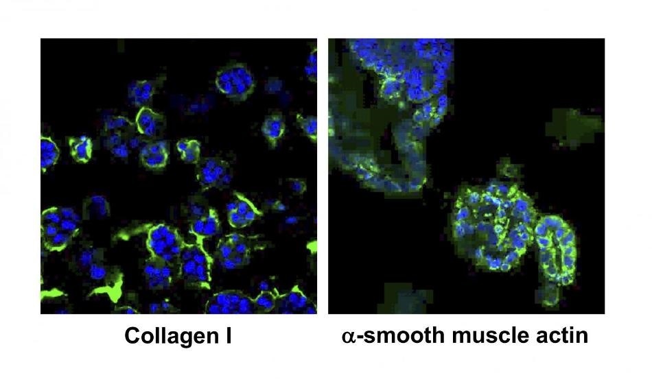
Expression of Collagen I protein in green indicates that fibrosis, one of the NASH characters, is formed in the NASH organoids. DAPI staining in blue shows nuclei of cells. Right panel: Expression of Smooth muscle actin protein in green indicates that hepatic stellate cells were activated in the NASH organoids. This also represents one of the NASH characters. Image Credit: T. Usui / Tokyo University of Agriculture and Technology.
The researchers developed the 3D culture by extracting cells from the liver tissues of NASH model mice. The team’s findings present an alternative option for creating drugs for patients suffering from NASH, detecting novel markers for early diagnosis, and a more improved interpretation of the progression of the disease.
The results of the study were published in the Biomaterials journal on January 27th, 2020.
In Japan, approximately 10 million people (that is, around 8% of the population) are believed to carry NASH or have a high risk for NASH. According to a ballpark estimate, NASH occurs in nearly 3% to 12% of adults in the United States.
Symptoms of NASH include fat deposition, fibrosis, and inflammation of the liver tissue. Incidentally, no medications are available for the treatment of NASH, making it a major social issue.
Hence, to identify the type of drugs that could cure NASH, the scientists used a technique in which experimental animals are fed by a NASH-inducing diet and then continuously administering drugs. But it is evident that the use of experimental animals for drug testing is not a viable solution.
To mimic NASH in a dish, we started three-dimensionally growing cells that were collected from liver tissues of NASH model mice. These cells successfully grew in a dish and formed mini-organs called organoids.”
Dr Tatsuya Usui, Senior Assistant Professor, Laboratory of Veterinary Pharmacology, Department of Veterinary Medicine, Faculty of Agriculture, Tokyo University of Agriculture and Technology
Dr. Usui is also the study’s corresponding author.
To produce NASH organoids in a dish, the researchers isolated cells from the liver of the NASH mice exhibiting three different disease stages—for example, the early stage (fatty liver), the middle stage (fatty liver), and the late-stage (advanced fibrosis).
The NASH organoids were analyzed using standard histology techniques, like oil red staining, HE staining, and Masson’s trichrome staining to inspect the production of oil in cells, the shapes of the cells, and the connective tissues, respectively. Apart from these methods, RNA-sequencing, quantitative PCR, and immunostaining were carried out to observe localization and the proportion of bio-markers.
After careful scientific analyzes, we found that these cells’ characters in the organoids were very similar to those of NASH liver tissues. Interestingly our NASH organoids also mimic characters of these stages. We therefore concluded that NASH was reproduced in a dish using the organoid culture method for the first time. We expect that drug discovery targeted for each stage can be done using these organoids.”
Dr Tatsuya Usui, Senior Assistant Professor, Laboratory of Veterinary Pharmacology, Department of Veterinary Medicine, Faculty of Agriculture Tokyo University of Agriculture and Technology
“In addition, our RNA-sequencing analysis found that several genes were elevated at all stages of organoids and some others were highly expressed at a specific state (patent application filed). So far, no effective diagnostic biomarkers have been found that accurately reflects the degree of progression of the NASH. Therefore, we expect that new reliable bio-markers for diagnosis can be identified using our NASH organoids,” Usui concluded.
Source:
Journal reference:
Elbadawy, M., et al. (2020) Efficacy of primary liver organoid culture from different stages of non-alcoholic steatohepatitis (NASH) mouse model. Biomaterials. doi.org/10.1016/j.biomaterials.2020.119823.