Below the surface of the ocean, a virus is stealing the metabolism of the most abundant organism present on Earth. That might be attractive to those who breathe above the ocean’s surface.

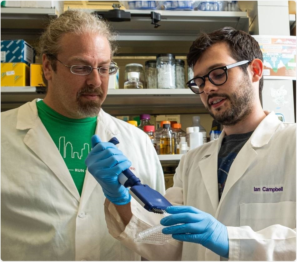
Rice University synthetic biologist Jonathan Silberg, left, and postdoctoral researcher Ian Campbell led a team that analyzed the role of ferredoxin proteins produced when viral phages alter electron transfer in ocean-dwelling, photosynthetic bacteria that produce oxygen and store carbon. Image Credit: Jeff Fitlow.
Researchers from Rice University examined the function of ferredoxin proteins that are created when phages modify the capacity of Prochlorococcus marinus to preserve carbon and offset the effect of greenhouse gas emerging from the consumption of fossil fuel.
The photosynthetic cyanobacteria P. marinus are mainly found in the tropics and subtropics where an estimated 1027 (an octillion) of the pathogens make use of sunlight to generate oxygen and collectively preserve 4 Gt of carbon every year. Some amount of this carbon serves as a critical feedstock for other marine organisms.
However, phages are their enemies. The virus reinforces itself by hijacking the energy produced by the bacteria from light and reprograms its victim’s genome to modify the way it transfers electrons.
According to Ian Campbell, a postdoctoral researcher at Rice University and the study’s lead author, P. marinus as well as its carbon-storing mechanism are responsive to temperature, so it is worth watching as climate change heats the oceans and extends its range.
The growth in the range of this organism in the oceans could increase the total carbon stored by these microbes. Alternatively, the viruses that infect these bacteria could alter carbon fixation and potentially prevent gigatons of carbon from being taken out of the air annually, according to one recent projection.”
Ian Campbell, Study Lead Author and Postdoctoral Researcher, Rice University
Campbell explained that the aim of this study was to analyze the various ways by which viruses communicate with their hosts. While doing so, the researchers found that the phage takes control of the flow of electrons in the host itself and rewires the metabolism of the bacteria.
Campbell added, “When the virus infects, it shuts down production of the bacterial proteins and replaces it with its own variants. I compare it to putting a different operating system on a computer.”
The scientists employed synthetic biology methods to mix and match the cyanobacterial proteins and phage to study their interactions. Rice biochemist George Phillips, who led a part of the study, determined the structure of a crucial cyanophage ferredoxin protein, for the first time.
A phage would usually go into a cell and kill everything. But Ian’s results suggest these phages are establishing a complex control mechanism. I wouldn’t say they’ve zombified their hosts, because they allow the cells to continue doing some of their own housekeeping. But they’re also plugging in their own ferredoxins, like power cables, to fine tune the electron flow.”
Jonathan Silberg, Study Lead Scientist and Synthetic Biologist, Rice University
Silberg is also the director of the Systems, Synthetic, and Physical Biology program at Rice University.
Campbell and his research team made use of synthetic biology tools, rather than working directly with P. marinus and cyanophages. They used these tools to reprogram relatively larger and better understood Escherichia coli bacteria to express genes that imitated the communications between the two.
Taking a phage and a cyanobacteria from the ocean and trying to study the biology, especially electron flow, would be really hard to do through classical biochemistry. Ian literally took partners from both the phage and the host, put them together by encoding their DNA in another cellular system, and was able to quickly develop some interesting results.”
Jonathan Silberg, Study Lead Scientist and Synthetic Biologist, Rice University
Silberg continued, “It’s an interesting application of synthetic biology to understand complex things that would otherwise be arduous to measure.”
The scientists suspect that the protein they modeled in Escherichia coli—the Prochlorococcus P-SSM2 phage ferredoxin—is not a novel concept.
“People knew phages encode different things that do electron transfer, but they didn’t know how to connect the wires between the phage and the host. They also didn’t know a lot about the phage’s evolution. The structure makes it clear this phage can be traced to specific ancestral proteins involved in photosynthesis,” Silberg concluded.
Source:
Journal reference:
Campbell, I. J., et al. (2020) Prochlorococcus phage ferredoxin: Structural characterization and electron transfer to cyanobacterial sulfite reductases. Journal of Biological Chemistry. doi.org/10.1074/jbc.RA120.013501.