In association with the Hubrecht Institute in The Netherlands, researchers from the University of Cambridge have devised a new model to analyze an early stage of human development through human embryonic stem cells.

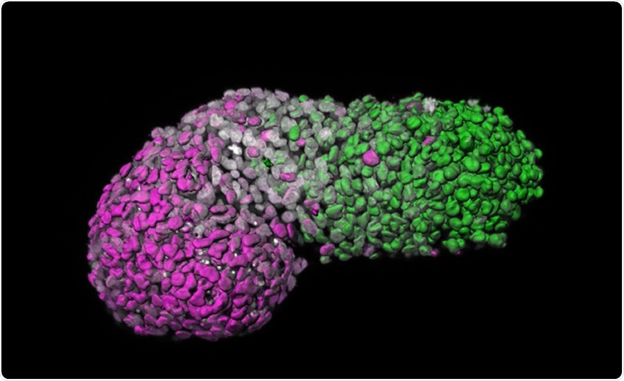
Image analysis of human gastruloid showing “anteroposterior” patterning. Green is the posterior part, similar to the tail-end of an embryo; magenta is the anterior part, similar to developing heart cells; grey marks DNA. Image Credit: Naomi Moris.
The new model mimics certain major elements of an embryo at about 18–21 days old and enables the scientists to visualize the processes fundamental to the formation of the human body blueprint that were never directly seen before. A better insight into these processes could expose the factors that are responsible for causing human birth defects and diseases and to create tests for these anomalies in pregnant women.
The blueprint, or body plan, of an organism, emerges through a process known as “gastrulation.” During this process, three distinct cellular layers are formed in the embryo that will subsequently give rise to all the major systems of the body: the endoderm will make the gut, ectoderm the nervous system, and mesoderm the muscles.
Gastrulation is usually known as the “black box” period of human development because as per legal restrictions, human embryos cannot be cultured in the laboratory beyond day 14 when the process begins. This limitation was fixed to fall at the phase where the embryo is no longer able to form a twin.
Several birth defects emerge during this period of “black box,” with causes including medications, infections, alcohol, and chemicals. A deeper understanding of human gastrulation could also shed new light on several medical problems, such as genetic disorders, miscarriage, and infertility.
Our model produces part of the blueprint of a human. It’s exciting to witness the developmental processes that until now have been hidden from view—and from study.”
Alfonso Martinez-Arias, Study Lead and Professor, Department of Genetics, University of Cambridge
A study published recently in the Nature journal demonstrates a technique of using human embryonic stem cells to produce a three-dimensional (3D) assembly of cells known as gastruloids. These cells differentiate into three different layers arranged in a way that mimics the early human body blueprint.
To culture gastruloids in the laboratory, specified numbers of human embryonic stem cells were transferred into small wells, where they developed into tight aggregates. Upon treating with chemical signals, lengthy gastruloids were observed to extend along a head to tail axis referred to as the anteroposterior axis, switching on genes in certain patterns along this axis that mirror the elements of a mammalian body blueprint.
Model organisms like zebrafish and mice have earlier allowed researchers to gain some understanding of human gastrulation. But these models may act differently to human embryos when the cells undergo differentiation.
Animal models can react differently to specific drugs: for example, the anti-morning sickness drug thalidomide cleared clinical trials after it was tested in mice but later caused severe birth defects in humans. Due to this reason, it is crucial to develop more improved models of human development.
Gastruloids lack the ability to develop into a fully mature embryo. They also lack brain cells or any other tissues required for implantation in the womb. This implies that they will not be able to develop beyond the very early stages of development, and thus conform to present ethical standards.
By observing the types of genes that were expressed in these human gastruloids after 72 hours of development, the scientists found an evident sign of the event that results in significant body structures, like cartilage, thoracic muscles, and bones, but they do not develop the brain cells.
The scientists were able to judge the corresponding human embryonic age of the gastruloids by evaluating them against the Carnegie Collection of Embryology. This official collection comprises a continuum of human embryos, such as daily growth across the first eight weeks. They imply that gastruloids partly mimic the 18- to 21-day-old human embryos.
This is a hugely exciting new model system, which will allow us to reveal and probe the processes of early human embryonic development in the lab for the first time. Our system is a first step towards modelling the emergence of the human body plan, and could prove useful for studying what happens when things go wrong, such as in birth defects.”
Naomi Moris, Study First Author, Department of Genetics, University of Cambridge
Produced from human stem cells, the novel “gastruloid” model holds a great potential to figure out the causes of birth defects and enhance the modeling of various diseases.
Source:
Journal reference:
Moris, N., et al. (2020) An in vitro model of early anteroposterior organization during human development. Nature. doi.org/10.1038/s41586-020-2383-9.