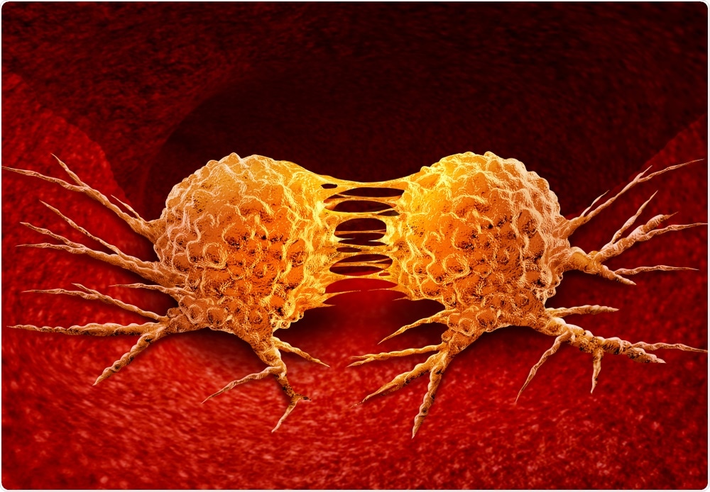
Metastasis - the development of tumor growth at a secondary site - is responsible for the majority of cancer-related deaths. It occurs when the primary tumor site sheds cancerous cells which are then circulated through the body via blood vessels or lymph nodes. These become seeds for eventual tumor growth at a secondary location in the body.

Image Credit: Lightspring/Shutterstock.com
Detection of these very rare cells, known as circulating tumor cells or CTCs, is important for early prognosis of serious disease as well as to monitor the effectiveness of treatment. Currently, there is only one method for CTC detection approved by the U.S. Food & Drug Administration (FDA), CellSearch, which is used to diagnose breast, colorectal and prostate cancer.
Results from a recent study - a collaboration between Lehigh University, Lehigh Valley Cancer Institute, and Pennsylvania State University - demonstrates the potential for a new method of detecting circulating tumor cells.
Unlike existing methods, which rely on an expensive and time-consuming process that involves labelling antibodies with fluorescence, this technique uses a powerful label-free detection method.
Developed by Yaling Liu, a faculty member in Lehigh's Department of Bioengineering and in the Department of Mechanical Engineering and Mechanics, in collaboration with Xiaolei Huang, faculty member in Penn State's College of Information Sciences and Technology, the technique applies a machine learning algorithm to bright field microscopy images of cells detected in patient blood samples containing white blood cells and CTCs.
The blood samples were drawn from participating patients undergoing treatment for stage 4 renal, or kidney, cancer at Lehigh Valley Hospital-Cedar Crest under the care of Suresh G. Nair, M.D., Physician in Chief at Lehigh Valley Cancer Institute.
The model yielded a high rate of accuracy: 88.6% overall accuracy on patient blood and 97% on cultured cells. The results have been published in Nature Scientific Reports in an article called "Label-free detection of rare circulating tumor cells by image analysis and machine learning."
In addition to Liu, Huang, and Dr. Nair, authors include three Lehigh PhD students Shen Weng, Yuyuan Zhou and Xiachen Qin .
Dr. Nair says Liu's innovative technique to isolate rare circulating cancer cells in a tube of blood - which can number as few as 15 cells in one billion - represents "a simpler, elegant and cost effective approach to monitoring patients on therapies such as immunotherapy and targeted therapy for cancer at the circulating cell level rather than scans such as CAT scans, which look for 100 million or more cells organized into a one centimeter tumor."
This study, though small, demonstrates that our method can achieve high accuracy on the identification of rare CTCs without the need for advanced devices or expert users, thus providing a faster and simpler way for counting and identifying CTCs. With more data becoming available in the future, the machine learning model can be further improved and serve as an accurate and easy-to-use tool for CTC analysis."
Liu
The method, he says, requires minimal data pre-processing and has an easy experimental setup. To arrive at the results, the team preprocessed the whole blood samples, capturing the bright field and fluorescent images of the cells.
They trained a deep learning model with cropped single-cell in bright field images and used the corresponding fluorescent images as ground truth labels. They also trained and tested a model with cultured cell lines as a comparison. The group then did the testing and summarized the statistical results of the trained model.
"We tuned the details of the model to reach better results until the outcome reached the state-of-the-art," says Liu.
They essentially ran two experiments: one was a comparison group operated on the white blood cells and cultured cancer cell lines, and the other operated on the white blood cells and patient CTCs. They expected the first group of experiments using the comparison group to work fine because of the large number of training datasets for cultured cells.
They used 1745 single-cell images and achieved an overall accuracy of 97.5%. The team did not expect the second group, from patient blood samples, to yield as high an accuracy rate as the first group because the training dataset was limited?based on 95 single-cell images as raw input.
"But when we applied the transfer learning technique with the pre-trained network, we were surprised by the improvement," says Liu. "We found that the machine learning model can identify CTCs with reasonable accuracy as good as 88%."
The blood samples were collected partially using a commercial enrichment kit and partially using a microfluidic device developed by Liu specifically to catch and release CTCs. He and his team continue to innovate in this area and are developing a device that combines optical image machine learning and acoustic sorting to automatically process the sample.
Dr. Nair and Liu, along with two Lehigh Valley Health Network Oncology Fellows Dr. Zach Wolfe and Dr. Saro Sarkisian, will continue to collaborate on the next steps. Among those is refining the technique to look at DNA mutation changes in the captured cells.
According to Liu, this will provide even more information to physicians and enable them to make treatment adjustments that improve health outcomes, including prolonging patients' lives.
The work in this study was partially supported by a National Institutes of Health (NIH) grant, a Pennsylvania Infrastructure Technology Alliance (PITA) grant, the Pennsylvania Commonwealth Universal Research Enhancement Program (CURE), and the Andy Derr Foundation for Kidney Cancer Research .