According to a study published in the Neuron journal in September 2020, immune cells play a surprising role in fine-tuning the neural circuits of the brain.

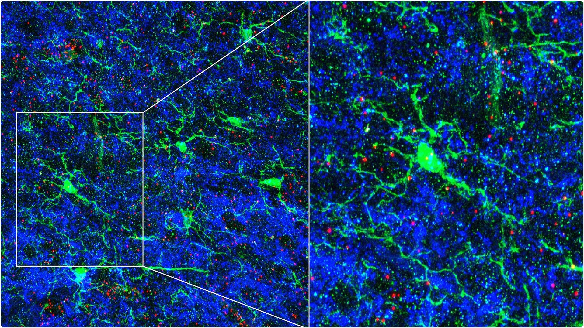
Microglia affects the density of neuronal connections. Microglial cells (stained in green), the immune cells of the mouse brain, send branches into areas with many neuronal connections (stained blue). Microglia makes an immune signal, or cytokine, called TWEAK at the ends of their branches (stained in red). Microglia that contain large amounts of TWEAK (are deeper red) contact fewer neuronal connections (seen as black areas without blue connections). Image Credit: Lucas Cheadle.
Called microglia, the immune cells residing there protect the brain against inflammation and infection and also support physically sculpt circuits in the developing brain. The latest study shows that microglia also guide neurons to alter their own connectivity in response to sensory cues.
Lucas Cheadle, an Assistant Professor from Cold Spring Harbor Laboratory (CSHL), identified this cellular communication while he was working as a postdoctoral researcher in Michael Greenberg’s laboratory. Greenberg is a neuroscientist from Harvard Medical School.
Cheadle’s study investigates the relationship between the external world and biology, and he is specifically keen to know how neural circuits are refined in response to sensory experience.
To a large extent, the general architecture and wiring of the brain is accomplished by birth. But it really requires this robust feedback from the environment to continue that maturation.”
Lucas Cheadle, Assistant Professor, Cold Spring Harbor Laboratory
Cheadle explained that when an animal interacts with its environment, certain neuronal connections are removed while others are reinforced. In humans, this kind of process continues for many years even after birth.
What's really important’s that during development, the right neurons connect with one another in the right way. We want to have tight control over how many connections there are and how strong the connections are. So that’s actually something that sensory experience is important for.”
Lucas Cheadle, Assistant Professor, Cold Spring Harbor Laboratory
Cheadle and collaborators tracked the relationship between synapses, or neurons, in a visual processing circuit in mice’s brain. Young mice require visual input at the correct time to develop brain pathways associated with vision.
However, if the mice lack visual input for a crucial period, the circuits release an excess number of synapses and the mice eventually end up with abnormal connections. The researchers also discovered that the circuits depended on microglia, which, with the correct visual stimuli, signaled adjacent neurons to prune a few synapses.
This effect on neural connectivity denotes a novel role for microglia in the healthy brain, and may help describe why the cells have been implicated in various neurodevelopmental disorders, including autism.
I think this study will be seen as a big breakthrough in our mechanistic understanding of how sensory experience and microglia coordinate the process of synaptic pruning that is critical for brain maturation in early life.”
Michael Greenberg, Neuroscientist, Harvard Medical School
At CSHL, Cheadle will study such an interaction in more detail, tracing the molecular signals that result in the disassembly of synapses and also the alterations that occur inside the microglia in response to environmental cues.
“The very idea that microglia are able to upregulate the expression of genes in response to visual experience in and of itself is fascinating for me, because these are immune cells,” Cheadle concluded.
Source:
Journal reference:
Cheadle, L., et al. (2020) Sensory Experience Engages Microglia to Shape Neural Connectivity through a Non-Phagocytic Mechanism. Neuron. doi.org/10.1016/j.neuron.2020.08.002.