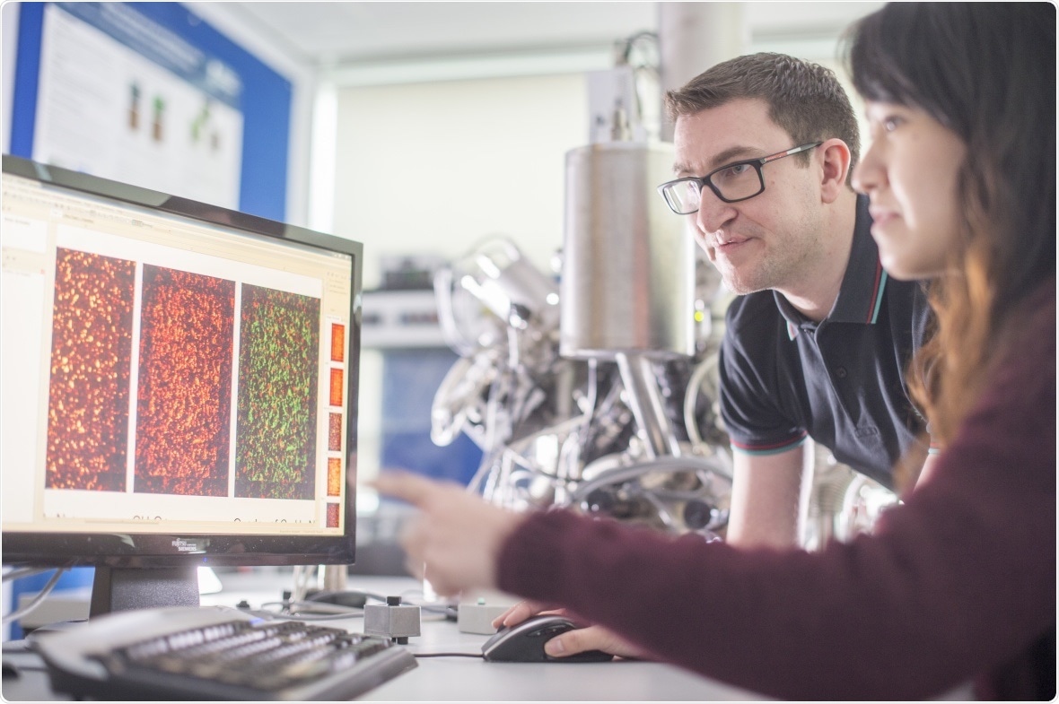
Researchers have defined a new protein imaging technique that may result in novel discoveries in disease, cell analysis, biological tissue, and more. The method may also prompt the development of novel biomaterials that can be used for advanced medical devices and drug delivery systems.

Image Credit: University of Nottingham.
A research team from the University of Nottingham, in association with the University of Birmingham and The National Physical Laboratory, has applied the next-generation three- dimensional (3D) OrbiSIMS instrument to enable the first matrix-free and label-free in situ assignment of intact proteins at surfaces without requiring major sample preparation.
The researchers’ study was recently published in the Nature Communications journal.
The University of Nottingham is the world’s first University to own a 3D OrbiSIMS instrument. It can now enable an unparalleled level of mass spectral molecular analysis for a variety of materials, including tissues, biological cells, and hard and soft matter.
Moreover, the facility in the University of Nottingham has high pressure freezing cryo-preparation amenities that allow researchers to preserve the biological specimens close to their native state as frozen-hydrated to balance the more commonly used but more disruptive freeze-drying and sample fixation.
When the high mass, surface sensitivity, and spatial resolution are integrated with a depth profiling sputtering beam, the OrbiSIMS instrument becomes a highly robust tool for 3D chemical analysis, as shown in this new study.
Dr. David Scurr from the School of Pharmacy at the University of Nottingham headed the new study and was supported by Anna Kotowska, a Ph.D. student.
The design and innovation of the next generation of biomaterials is underpinned by the ability to accurately characterise biological tissue and materials. The challenge for scientists in this area has been unpicking the chemical complexity of such systems.”
Dr David Scurr, School of Pharmacy, University of Nottingham
Dr. Scurr continued, “This approach to protein analysis has been demonstrated using extreme examples to illustrate its sensitivity and specificity by chemically mapping a protein monolayer (protein biochip) and distribution of specific protein in human skin (complex multi-layered biological system) respectively.”
With the ability to chemically map proteins in this way we are a step closer to being able to understand fundamental biological processes and develop more effective systems for targeting drugs and providing coatings for medical devices.”
Dr David Scurr, School of Pharmacy, University of Nottingham
The researchers from the University of Nottingham have already performed biomaterials studies to develop a new kind of urinary catheter in association with Camstent Ltd. The catheter is coated in a bacteria-resistant material found by investigators from the University of Nottingham.
Morgan Alexander, a Professor and Director of the EPSRC Programme Grant in Next Generation Biomaterials Discovery and the 3D OrbiSIMS facility, stated “The research we are now able to do using this instrument is paving the way for step changes in how materials can be used in medicine to better treat disease and illness.”
“The Catheter coating we have developed in partnership with Camstent has gone all the way from the discovery of a new class of materials that no one could have predicted all the way to clinical trials and is a great example of the application of this type of research,” Alexander added.
With these new capabilities to characterise proteins at surfaces comes also new exciting opportunities to engineer functional materials with predictable protein interactions for biosensor technology.”
Paula Mendes, Professor of Advanced Materials and Nanotechnology, University of Birmingham
Source:
Journal reference:
Kotowska, A. M., et al. (2020) Protein identification by 3D OrbiSIMS to facilitate in situ imaging and depth profiling. Nature Communications. doi.org/10.1038/s41467-020-19445-x.