The human body experiences ten quadrillion cell divisions during an individual’s lifetime. Such a biological process is crucial to form and sustain organs and tissues inside the body.

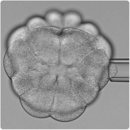
Measuring the stiffness of dividing cells. An ascidian consisting of 24 cells. The stiffness of the cells preparing for cell division is measured with a micropipette. Image Credit: © Benoit Godard/ Institute of Science and Technology Austria.
Professor Carl-Philipp Heisenberg and his group from the Institute of Science and Technology Austria have now discovered how the division process is influenced by a mechanical tension from surrounding tissue.
The researchers have published the results of their study in the Developmental Cell journal. The study shows an entirely new impact on cell division and may also be significant for tumor research.
A parent cell splits into two daughter cells during cell division. The new role and function of the daughter cells rely on the orientation of the division plane. The shape of the mother cell is a vital factor that directs this orientation of cell division.
In association with the McDougal laboratory at Sorbonne University, Professor Heisenberg and Benoit Godard, a Postdoc in Heisenberg’s laboratory, discovered that the dividing cell softens and, therefore, becomes deformable by mechanical tension arising from adjacent cells. This shows how tissue tension affects the orientation of cell division.
The neighboring tissue pulls and deforms the soft dividing cells. Consequently, this alters its cell division orientation and, therefore, the fate of its daughters.”
Carl-Philipp Heisenberg, Professor, Institute of Science and Technology Austria
A new model organism
It is difficult to study cellular interactions inside a tissue. Because of the involvement of a huge number of cells, it is hard to disentangle the cause and effect. Hence, Professor Heisenberg employed a special model organism.
Small marine invertebrates, called ascidians, develop almost identical to vertebrates but possess only a minimal number of cells. In addition, the fate of the cell is set quite early. With a predetermined development and fewer cells, ascidians enable researchers to study the tissue dynamics more accurately.
Observing dynamics cell shaping
For over a century, it has been known that cells are inclined to divide perpendicular to their long axis. In the past, cells experiencing division were believed to be stiff and, hence, insensitive to mechanical forces from their surroundings, influencing their shape and thereby the orientation of cell division.
Contrary to these beliefs, Professor Heisenberg and his group have now identified that dividing cells can turn softer and, hence, can be deformed by extrinsic forces.
We had preliminary and indirect results suggesting that dividing cells become soft, but this was opposite to the literature on cell division. Thus, when I conducted the biophysical measurement of cell stiffness and saw the softening occurring, it was the greatest moment in my scientific career.”
Benoit Godard, Postdoc, Institute of Science and Technology Austria
The researchers carried out a three-dimensional scan of the shapes of cells in the developing ascidian embryos and integrated it with biophysical measurements of cell stiffness. This process allowed them to learn that the dividing cells turn softer and thus become deformed in response to forces from adjacent cells.
“We wanted to identify the governing factors of cell division. We found that cells become soft when undergoing division and that this allows extrinsic mechanical forces to influence the shape and thus division orientation of the dividing cell.”
Carl-Philipp Heisenberg, Professor, Institute of Science and Technology Austria
The new study paves the way to innovative fundamental studies and may also be relevant for clinical research. For instance, tumors modify tissue tension, which again probably influences the rate and division orientation of tumor cells. These outcomes help describe the malformation of tissues and may allow the externally controlled formation of tissues.
Deformation of soft dividing cells by tissue tension
An ascidian embryo at the 32-cell stages. The asterisks mark soft cells at the bottom, which enter cell division and orient their division plane perpendicular to the non-dividing cells at the top by which they are pulled and deformed. Video Credit: Benoit Godard/Institute of Science and Technology Austria.
Source:
Journal reference:
Godard, B. G., et al. (2020) Apical Relaxation during Mitotic Rounding Promotes Tension-Oriented Cell Division. Developmental Cell. doi.org/10.1016/j.devcel.2020.10.016.