A new, open-source tool enables non-experts to interpret microscopy images using artificial intelligence. The tool, which was created at Finland’s Åbo Akademi University and Portugal’s Instituto Gulbenkian de Ciência, would be extremely useful in analysis and diagnostics using modern microscopes.

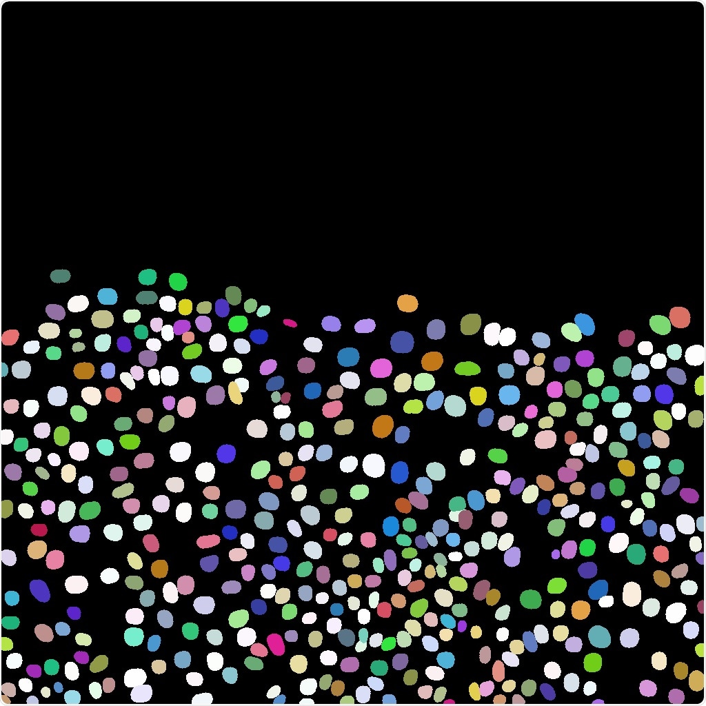
Example illustrating how AI via ZeroCostDL4Mic can be used to detect the nucleus of cancer cells from microscopy images. Upper picture: Original microscopy image. Lower picture: Image where each detected cancer cell has a different color. Image Credit: Guillaume Jacquemet.
Artificial intelligence, or AI, software has been completely transforming the way microscopy images are interpreted. For example, AI can be used to identify features in images (that is, tumors in biopsy samples) or to increase image quality by eliminating unnecessary noise. Non-experts, on the other hand, continue to struggle with AI technologies.
Researchers have outlined a tool named ZeroCostDL4Mic in their article titled “Democratizing deep learning for microscopy with ZeroCostDL4Mic,” which was published in Nature Communications on April 15th, 2021.
The key novelty is that ZeroCostDL4Mic runs in the cloud for free and does not require users to have any coding experience or advanced computational skills. Effectively, it runs on any computer that has a web browser.”
Guillaume Jacquemet, Senior Researcher in Cell Biology, Åbo Akademi University
For the past four centuries, microscopes have enabled humans to view objects that would otherwise be too small to see with the naked eye. Microscopy is now a leading technology used globally not just for research but also for diagnostics.
Modern microscopes are directly linked to digital cameras, enabling the capture of hundreds to thousands of images per sample. To obtain useful data from these images, they must be processed with a computer, which is a mammoth task.
Jacquemet and his collaborators used AI to teach a machine to do the work to assist with the volume of images. ZeroCostDL4Mic is actually a collection of self-explanatory notebooks for Google Colab, with a user-friendly graphical user interface.
We believe that ZeroCostDL4Mic will acts as ‘a gateway drug’ for AI, luring users to explore these new technologies that will transform biomedical research and diagnostics in the decades to come.”
Guillaume Jacquemet, Senior Researcher in Cell Biology, Åbo Akademi University
Source:
Journal reference:
Chamier, L. V., et al. (2021) Democratising deep learning for microscopy with ZeroCostDL4Mic. Nature Communications. doi.org/10.1038/s41467-021-22518-0.