COVID-19 pandemic has emphasized the need for techniques to recognize repurposed or new drugs as antivirals. Scientists from the Uppsala University and Karolinska Institutet are exploring a novel screening approach that concentrates on the recognition of virus-specific morphological changes in virus-infected cells.

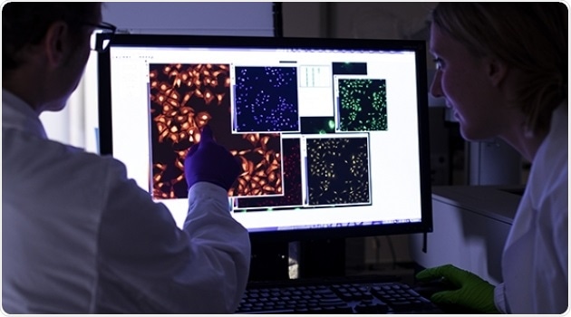
Jordi Carreras-Puigvert and Jonne Rietdijk studying cell images being acquired by a high-content microscope. Image Credit: Uppsala University.
The current approach concentrates on determining the changes (morphological profiles) that the virus creates on the infected cells by employing a revised version of the Cell Painting protocol, a recognized assay that utilizes a cocktail of fluorescent reagents to stain numerous cellular compartments. These morphological profiles are later utilized as a basis to identify drugs that can reverse the virus-induced effects.
In a single assay combining Cell Painting with antibody-based detection of viral infection at a single cell level, the scientists were able to substantiate the antiviral effect of known reference drugs along with identifying new compounds as potential antivirals.
The process involves both image and data analysis pipelines using CellProfiler, a well-known image analysis software, which the scientists have made public to enable the use and spread of the new method.
Most of the approaches involved in the discovery of antiviral drugs available today focus on the effects of similar drugs on a given virus, its enzymatic activity, or constituent proteins all the while ignoring the consequences for host cells.
This is a problem, as potential toxicity impacting the overall physiology of host cells may mask the effects of both viral infection and drug candidates. With our method, on the other hand, we are able to assess the general health of host cells, and in parallel identify antiviral properties of compounds.”
Jordi Carreras-Puigvert, Study Senior Author and Lecturer, Pharmaceutical Bioinformatics Group, Department of Pharmaceutical Biosciences, Uppsala University
According to Jonne Rietdijk, “The reasoning behind our approach was to obtain unbiased morphological profiles in the context of the host cell to study viral infection and compound treatment in a single assay.”
We modified the Cell Painting protocol, combining it with a virus-specific antibody staining. This enabled us to select the virus-infected cells with high precision and even relate the morphological profiles with the viral protein levels in each cell.”
Jonne Rietdijk, Study First Author and PhD Student, Pharmaceutical Bioinformatics Group, Department of Pharmaceutical Biosciences, Uppsala University
The scientists demonstrate that their approach can efficiently capture virus-induced phenotypic signatures of human lung fibroblasts infected with the human coronavirus. They also showed that the process can be employed in phenotypic drug screening using a panel of nine host- and virus-targeting antivirals and that treatment with effective antiviral compounds reversed the morphological profile of the host cells toward a non-infected state.
“By only doing minor adjustments to the image analysis pipeline that we provide, we anticipate that our untargeted approach, will enable other applications using diverse (human-derived) cell lines, as well as different viruses,” says Jonne Rietdijk.
Jordi Carreras-Puigvert adds, “There are two main uses that we see for the method, one is the screening for already medically available drugs that could be repurposed as antivirals. The second one is the screening for actual novel compounds.”
Since we create compound-specific signatures, we can then compare these to a set of signatures extracted from compounds with known mode of action, thereby potentially identifying the target of a given novel compound, in this case an antiviral.”
Jordi Carreras-Puigvert, Study Senior Author and Lecturer, Pharmaceutical Bioinformatics Group, Department of Pharmaceutical Biosciences, Uppsala University
Source:
Journal reference:
Rietdijk, J., et al. (2021) A phenomics approach for antiviral drug discovery. BMC Biology. doi.org/10.1186/s12915-021-01086-1.