Micronuclei—small nucleus-like structures within cells—are generally linked with tumors. Recently, scientists from Tsukuba, Japan, created an automated computer program that can precisely and reproducibly count micronuclei from microscope images, which would accelerate the speed and accuracy of micronuclei research.

Image Credit: Image by Kateryna Kon/Shutterstock.
Recent research from the University of Tsukuba declared their novel MATLAB-based program, called CAMDi (Calculating Automatic Micronuclei Distinction), which automatically counts the micronuclei from images of stained cells. Micronuclei can be stained similar to regular nuclei; however, they are distinguished from nuclei by their very small size.
But pinpointing is tougher as automatic systems for counting micronuclei have conventionally employed images taken from just one level of tissue. To better understand its importance, one can imagine slicing a cross-section through a ball fixed in space.
When the cut occurs close to the bottom or top areas of the ball, the size of the cross-section will be smaller than a cut close to the center—hence, a cross-section adjacent to the periphery of a nucleus can possibly be mistaken for a micronucleus.
To address this issue, scientists from the University of Tsukuba took photos at various levels through cells or tissue and developed a program apt for evaluating the resulting three-dimensional information. The scientists, in this regard, assured that what the program counted as micronuclei were, in fact, micronuclei.
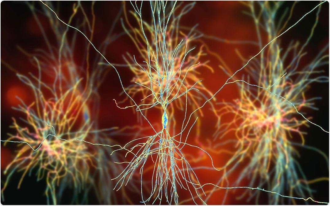
They later employed this program to search micronuclei in mouse neurons and analyzed the impacts of neuroinflammation on micronucleus numbers.
A link has been reported between inflammation and micronuclei in cancer cells. We decided to test whether neuroinflammation in the brain might affect the numbers of micronuclei in neurons.”
Dr Fuminori Tsuruta, Study Corresponding Author, University of Tsukuba
To achieve this, the scientists first introduced inflammatory factors into mouse neurons grown in culture; however, they observed no changes in micronuclei number with their CAMDi program.
However, when the mice were given injections of lipopolysaccharides, which induced the inflammatory cells in the hippocampal region to become activated, there was a rise in micronuclei in the hippocampal neurons.
These results were surprising. They suggest that the formation of micronuclei in neurons is induced by inflammatory responses from nearby cells.”
Dr Fuminori Tsuruta, Study Corresponding Author, University of Tsukuba
As micronuclei are markers of a range of pathologies, the designing of this novel computer program can be vital for pathological diagnoses and the tracking of treatment responses. CAMDi is anticipated to enhance the research into micronuclei—to provide a thorough knowledge of their formation and the roles they play in disease.
Source:
Journal reference:
Yano, S., et al. (2021) A MATLAB-based program for three-dimensional quantitative analysis of micronuclei reveals that neuroinflammation induces micronuclei formation in the brain. Scientific Reports. doi.org/10.1038/s41598-021-97640-6.