Amyloidosis, a multifaceted disease group results due to the misfolded “amyloid” protein deposits in different tissues. Amyloid protein is very distinct when compared to other native proteins as it possesses a cross-β-sheet structure. The structure provides unique properties like resistance to digestive proteinases and transmissibility through ingestion.

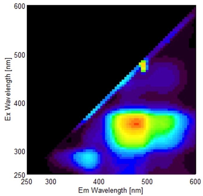
Fluorescence fingerprint of amyloid-containing bovine liver homogenate. Image Credit: Tomoaki Murakami/ Tokyo University of Agriculture and Technology.
Amyloidosis is presently diagnosed by employing time-consuming histopathological techniques. But these techniques require specific expertise for correct diagnosis through observation of specimens, not to mention the limitations. In the current research, scientists have effectively tested a novel process to identify AA amyloidosis in cattle intended to overcome the drawbacks of conventional detection.
The scientists from the Tokyo University of Agriculture and Technology (TUAT) and Konica Minolta Inc., Japan, jointly published their results on October 5th, 2021, in the Journal of Veterinary Diagnostic Investigation.
Actually, when we necropsied samples from chickens and quails, we found that the amyloid protein was visible in orange color with the naked eye. This observation led us to hypothesize that amyloid has some optical properties that normal proteins lack. On the other hand, we noticed that the bovine amyloid was not colored, so we came up with the idea that it might be fluorescent.”
Tomoaki Murakami DVM, PhD, Study Corresponding Author and Associate Professor, Laboratory of Veterinary Toxicology, Cooperative Department of Veterinary Medicine, Tokyo University of Agriculture and Technology
Numerous reports of intrinsic fluorescence of amyloid prevail. However, most of them employed amyloid fibrils artificially created in vitro. Moreover, there exists only a few researches on amyloid derived from living tissues. In the current research, the researchers experimented with their hypothesis on the qualities of bovine amyloid.
The researchers employed fluorescence fingerprint analysis on AA amyloid extracted from bovine livers. They identified that AA amyloid possessed a specific fluorescence fingerprint pattern. The researchers, through their study, created a quick and straightforward process for identifying amyloid in bovine liver homogenates.
Fluorescence fingerprint analysis is used for the analysis of food ingredients and other molecules, but surprisingly very few studies have used this technology in the area of clinical pathology. Fluorescence fingerprint analysis does not require any particular pre-treatment, hence the samples can be swiftly processed. Our detection method will likely lead to the development of more efficient and the rapid diagnostical tools for amyloidosis in the future.”
Tomoaki Murakami DVM, PhD, Study Corresponding Author and Associate Professor, Laboratory of Veterinary Toxicology, Cooperative Department of Veterinary Medicine, Tokyo University of Agriculture and Technology
Source:
Journal reference:
Ujike, N., et al. (2021) Intrinsic fluorescence–based label-free detection of bovine amyloid A amyloidosis. Journal of Veterinary Diagnostic Investigation. doi.org/10.1177/10406387211049217.