The brain has the potential to change the way neurons communicate with one another. That is, among other things, how it prevents out-of-control brain activity. Scientists from the University Hospital Bonn and an Australian team have discovered a mechanism that plays a key role in this.
This technique changes the synaptic coupling of neurons in cultured cells, affecting stimulus transmission and processing. Diseases including epilepsy, schizophrenia, and autism may arise if it is disrupted. The findings have been reported in Cell Reports.
In the human brain, around 100 billion nerve cells conduct their functions. Each of these neurons has 1,000 connections with other neurons on average. Information is transferred between nerve cells at these so-called synapses.

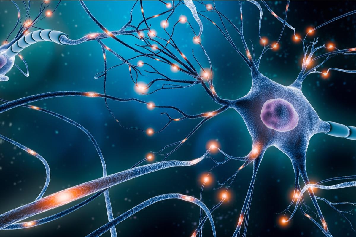
Image Credit: MattLphotography/Shutterstock.com
Synapses, on the other hand, are considerably more than just wires. Their structure already reflects this: They are made up of a presynapse, which is a type of transmitter, and a postsynapse, which is a receiver structure. The synaptic cleft is located between them. This is a relatively little space. Nonetheless, it makes it difficult for electrical impulses to be transmitted. Instead, the neurons communicate with each other across the gap by shouting information.
Incoming voltage pulses cause the presynapse to release particular neurotransmitters for this function. On the postsynaptic side, they traverse the synaptic cleft and attach to certain “antennae.” They also create electrical pulses in the receiver cell as a result of this.
However, the amount of neurotransmitter released by the presynapse and the extent to which the postsynapse responds to it are strictly regulated in the brain.”
Dr Susanne Schoch McGovern, Professor, Department of Neuropathology, University Hospital Bonn
Sophisticated control mechanisms
During learning, for example, specific synapses become stronger, allowing even a mild electrical stimulus from the transmitter neuron to generate a robust reaction in the receiver cell. Synapses that are rarely utilized, on the other hand, degrade. Furthermore, sophisticated regulatory systems prohibit the brain’s electrical activity from extending too far or, conversely, going away too soon.
We also speak of synaptic homeostasis, it ensures that brain activity is always within a healthy range.”
Dr Dirk Dietrich, Professor, Department of Neurosurgery, University Hospital Bonn
The mechanisms that keep this equilibrium in place, however, are still partially known. Homeostatic plasticity is a method through which the brain adjusts to long-term changes in neuronal activity. “We have now shown that a protein called RIM1 plays a key role in this process,” adds Schoch McGovern. RIM1 is concentrated in the presynapse’s “active zone,” which is where neurotransmitters are produced.
RIM1 is made up of a huge number of contiguous amino acids, much like any other protein. Scientists have previously discovered that an enzyme links some of these amino acids to a chemical molecule called a phosphate group. The presynapse can produce more or fewer neurotransmitters depending on which amino acid is changed in this way. The phosphate groups serve as the synapses’ “memory,” allowing them to recall their current activity level.
Dietrich states, “In the presynapse, transmitter-filled vesicles stand ready to be fired like the arrows of a taut bow. As soon as a voltage pulse comes in, they are released at lightning speed. Phosphorylation changes the number of these vesicles.”
Synapse calls with louder voice
If the presynapse can “fire” multiple vesicles as a result, its signal across the synaptic cleft gets louder. If on either hand, the number of vesicles drops dramatically as a result of changes in the RIM1 phosphorylation state, the call is hardly audible. “Which effect occurs depends on the phosphorylated amino acid,” explains Schoch McGovern’s study group’s Dr Johannes Alexander Müller. Alexander Müller also shares co-lead authorship of the paper with colleague Dr Julia Betzin.
This suggests that RIM1 may allow the brain to fine-tune the activity of individual synapses. The enzyme SRPK2 also plays an important function in RIM1: it binds the phosphate groups to the amino acids. Other participants, such as an enzyme that eliminate the phosphate groups—again if necessary, are also present.
We assume that there is a whole network of enzymes that act on RIM1 and that these enzymes also control each other’s activity.”
Dr Dirk Dietrich, Professor, Department of Neurosurgery, University Hospital Bonn
The synaptic equilibrium is extremely important; if it is interrupted, diseases like epilepsy, as well as schizophrenia and autism, may occur. RIM1 genetic information is frequently changed in patients with certain psychiatric disorders, which is interesting. This might indicate that the RIM1 protein in them is less effective.
“We now want to further elucidate these relationships. Perhaps new therapeutic options for these diseases will emerge from our findings in the long term, although there is certainly a long way to go before that happens,” Schoch McGovern, who is a member of the Transdisciplinary Research Area “Life and Health,” concluded.
Source:
Journal reference:
Müller, J. A., et al. (2022) A presynaptic phosphosignaling hub for lasting homeostatic plasticity. Cell Reports. doi.org/10.1016/j.celrep.2022.110696.