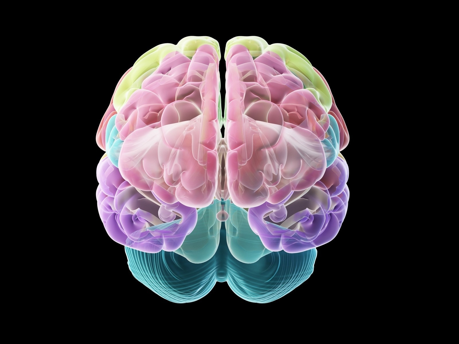
Researchers at Cedars-Sinai have produced an unprecedented number of bio-realistic and intricate computer models of individual brain cells.
 New research from Cedars-Sinai: Investigators have created bio-realistic and complex computer models of individual brain cells. Image Credit: Getty Images
They describe how these models could someday provide answers to issues regarding neurological illnesses and even human intelligence that cannot be investigated through biological trials in their research, which was just published in the peer-reviewed journal Cell Reports.
These models capture the shape, timing, and speed of the electrical signals that neurons fire to communicate with each other, which is considered the basis of brain function. This lets us replicate brain activity at the single-cell level.”
Costas Anastassiou, PhD, Study Senior Author and Research Scientist, Neurosurgery, Cedars-Sinai
For the first time, the models provide a comprehensive picture of the electrical, genetic, and biological activity of single neurons by combining data sets from several types of laboratory research. According to Anastassiou, the models can be used to evaluate theories that would necessitate conducting dozens of tests in the lab.
Imagine that you wanted to investigate how 50 different genes affect a cell’s biological processes. You would need to create a separate experiment to ‘knock out’ each gene and see what happens. With our computer models, we can change the expression level of genes at will for as many as we like and predict what will happen.”
Costas Anastassiou, PhD, Study Senior Author and Research Scientist, Neurosurgery, Cedars-Sinai
The cellular models also give researchers complete control over the parameters of their experiments, which is another benefit.
“This opens the possibility of establishing that one parameter, such as a protein expressed by a neuron, causes a change in the activity of that cell that eventually leads to a disease condition, such as epileptic seizures. In the lab, investigators can often show an association, but it is difficult to prove a cause,” Anastassiou said.
“In laboratory experiments, the researcher doesn’t control everything. Biology controls a lot. But in a computational simulation, all the parameters are under the creator’s control. In a model, I can change one parameter and see how it affects another, something that is very hard to do in a biological experiment,” Anastassiou added.
Using two different sets of data on the mouse primary visual cortex, the region of the brain that controls information from the eyes, Anastassiou and his group from the Anastassiou Lab—members of the Departments of Neurology and Neurosurgery, the Board of Governors Regenerative Medicine Institute, and the Center for Neural Science and Medicine at Cedars-Sinai—developed their models.
Tens of thousands of single cells’ complete genomic profiles were revealed in the first data collection. The second connected 230 cells from the same brain region’s electrical responses and physical features. The researchers combined these two datasets, using machine learning to produce bio-realistic models of 9,200 single neurons and their electrical activity.
This work represents a significant advancement in high-performance computing. It also gives researchers the ability to search for relationships within and between cell types and to glean a deeper understanding of the function of cell types in the brain.”
Keith L. Black, MD, Chair, Neurosurgery, Cedars-Sinai
Black was also the Ruth and Lawrence Harvey Chair in Neuroscience at Cedars-Sinai.
The Allen Institute for Brain Science in Seattle, which also contributed data, worked on the project alongside us.
“This work led by Dr. Anastassiou fits in well with Cedars-Sinai’s dedication to bringing together mathematics, statistics, and computer science with technology to address all the important questions in biomedical research and healthcare,” said Jason Moore, PhD, chair of the Department of Computational Biomedicine.
“Ultimately, this computational direction will help us understand the deepest mysteries of the human brain.”
To research human brain function and disease, Anastassiou and his team are now developing computer models of human cells.