Reviewed by Danielle Ellis, B.Sc.Jan 6 2023
Although the human body is superficially symmetric along the left–right axis, most internal organs, including the heart, lungs, liver, stomach, and brain, have noteworthy left–right asymmetries in their structure and placement.

Image Credit: SciePro/Shutterstock.com
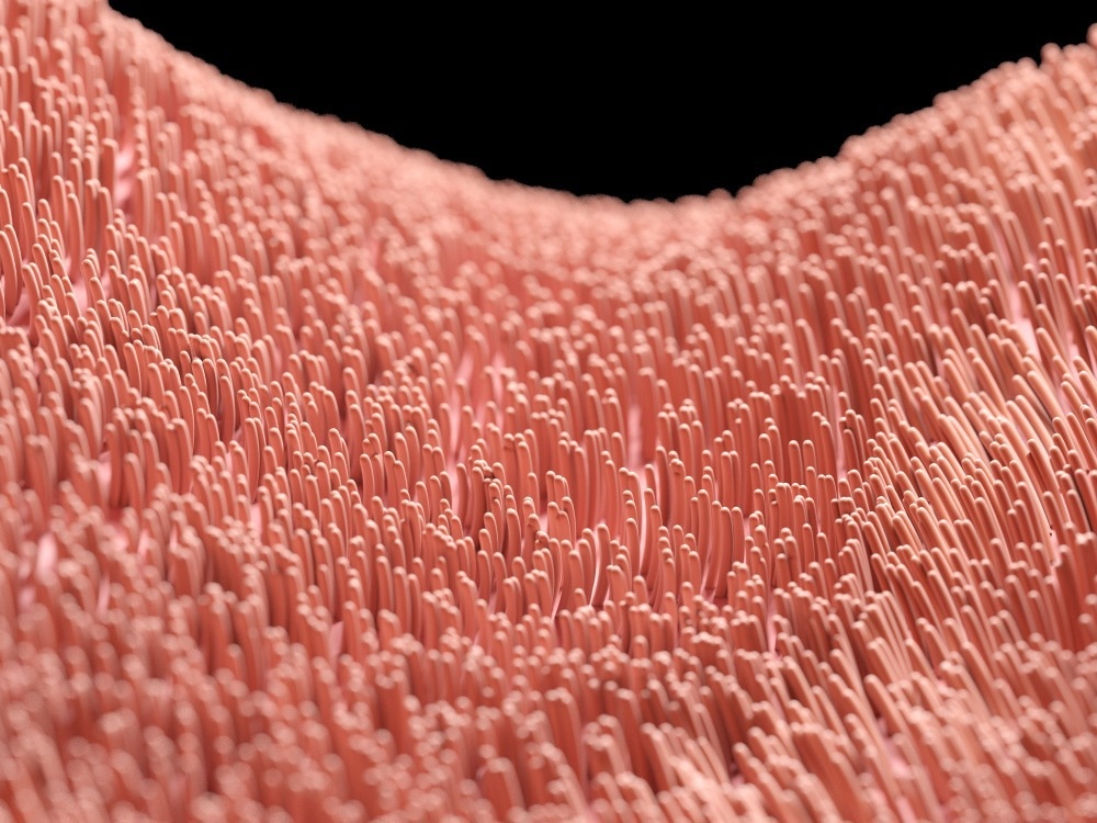
A tiny cluster of cells called the left–right organizer is known to establish left–right asymmetry during early development. Motile cilia, hair-like structures on cell surfaces, beat rapidly within this organizer to generate a leftward directional flow of extracellular fluid, the first outward sign of a left–right difference.
This early flow has been demonstrated to be crucial to distinguishing right from left; nevertheless, it is unknown how this flow is detected and translated into left–right asymmetry.
New research headed by MGH investigators reveals that cilia in the organizer not only operate as flow creators but also as sensors for the biomechanical forces produced by the flow to shape the growing embryo’s left–right body plan.
The research was published in the journal Science.
Nearly 25 years of work by numerous groups have shown that cilia and flow in the organizer are absolutely essential for establishing body left–right asymmetry. But we haven’t had the right tools or techniques to definitively study how this all works.”
Shiaulou Yuan PhD, Investigator, Cardiovascular Research Center, Massachusetts General Hospital
Shiaulou Yuan is also an assistant professor of medicine at Harvard Medical School and senior author of the study
To tackle this issue, the scientists used zebrafish as a model for left–right development, as well as a novel optical toolkit that included custom-built microscopy and machine learning analysis.
Their technique was exceptional in that they built and used optical tweezers—a biophysical tool that utilizes light to hold and move small objects akin to a tractor beam—for the first time, allowing precise mechanical force delivery onto cilia in an entire, living animal.
The scientists found that cilia are cell-surface mechanosensors that are essential for left–right asymmetry of the developing body and organs like the heart using these methods.
They demonstrated that a subset of organizer cilia sense and translate flow forces into calcium signals that regulate left–right development in zebrafish by employing optical tweezers to apply mechanical force to cilia in the left–right organizer.
Left–right asymmetry defects are linked to a variety of human disorders, including heterotaxy syndrome, primary ciliary dyskinesia, and congenital heart disease.
The knowledge gleaned from this study not only advances our understanding of the fundamental cellular processes that govern the development of the human body, they may also open new avenues for the development of novel diagnostics of these disorders. Additionally, this work may pave the way for targeted therapies on cilia signaling and mechanosensing to improve outcomes.”
Shiaulou Yuan PhD, Investigator, Cardiovascular Research Center, Massachusetts General Hospital
Yuan and his coworkers are still researching the molecular mechanisms governing cilia force sensing. They are also working on new ways for visualizing and manipulating cilia signaling, with the long-term goal of generating novel tools for the treatment of cilia-related disorders.
These results, and the tools that made it possible, have provided a new window into the developmental patterning of the embryo, and also opened Pandora’s box. It reminds us that we have so much more to learn about how cilia signaling and mechanobiology impact development and disease.”
Scott E. Fraser, Study Co-Author and Provost Professor, Biology and Bioengineering, University of Southern California
Source:
Journal reference:
Djenoune, L., et al. (2023). Cilia function as calcium-mediated mechanosensors that instruct left–right asymmetry. Science. doi.org/10.1126/science.abq7317.